Clinical Summary
A 35-year-old woman presents with a 2.5 cm, polypoid mobile mass attached to the left posterior vaginal wall midway between the cervix and introitus. The preoperative clinical diagnosis is vaginal cyst. On sectioning, a well-circumscribed nodule is noted with a smooth, glistening pale-pink to yellow surface devoid of hemorrhage or necrosis. By immunohistochemistry, the tumor cells are positive for CD34, ER and PR and negative for desmin, smooth muscle actin, and S100.
Master List
- Aggressive angiomyxoma
- Cellular angiofibroma
- Low-grade fibromyxoid sarcoma
- Myxoid liposarcoma
- Myxoid neurofibroma
Archive Case and Diagnosis
This case first appeared as Performance Improvement Program in Surgical Pathology (PIP) 2015, case 04, and is cellular angiofibroma.
Criteria for Diagnosis and Comments
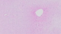
Histologic sections demonstrate a well-circumscribed but unencapsulated submucosal nodule directly abutting the mucosa. The lesion shows zones of loosely organized oval, spindle to stellate cells embedded in a pale, edematous stroma alternating with relatively densely cellular areas in which the cells are separated by wispy collagen strands and bundles. There is some variability from slide to slide and not all slides contain areas with a cellular stroma. Cells have opened chromatin, inconspicuous nucleoli, with indistinct delicate pale to eosinophilic cytoplasm. There is a mild degree of cytomorphologic variability but no overt pleomorphism or atypia. Mitotic figures are not easily found. Rare multinucleated cells can be identified. There are numerous small to medium-sized vessels evenly distributed throughout the lesion, many of which are hyalinized. More dilated and ectatic vessels are focally present, especially at the periphery. The lesional cells are positive for CD34 by immunohistochemistry but negative for desmin and S100. Taken together, the gross, histologic and immunohistochemical features are consistent with cellular angiofibroma.
Cellular angiofibroma (CAF) is an uncommon benign mesenchymal neoplasm of the vulvovaginal and inguinoscrotal region in women and men. Some authors consider this tumor to be synonymous with angiomyofibroblastoma while others regard them as distinct entities. Rare cases occurring at extra-genital sites like the eye and knee are described in the literature. While most cases arise in the subcutis, about 10% are dermally based. The lesion occurs most commonly in women between 40 to 50 years of age while men are affected at an older age (60-70 years); however, CAFs have been described in a wide age group (20 to 80 years). Clinically, CAFs may present with mild swelling and tenderness but most frequently as an asymptomatic polypoid or nodular mass of variable duration. Most patients seek medical treatment within a few weeks of noticing the lesion. In women, the most common clinical impression is that of a Bartholin gland or labial cyst with average size of 3 cm. Similarly, CAFs in men most often present as a painless, slowing enlarging mass in the scrotal/paratesticular region or inguinal/groin area. They are often found in the subcutis and on average tend to be somewhat larger (7 cm) than those typically seen in women. Rarely, CAFs may reach very large sizes. A notable example is a 25 cm CAF removed from the retroperitoneum of a 60-year-old man. Grossly, CAFs are well circumscribed and can be surrounded by a thin, compressed fibrous pseudocapsule. The cut surface is white-tan to grayish with solid to partly gelatinous or cystic areas. Foci of hemorrhage and necrosis are exceptionally rare. To date, clinical follow-up indicates a benign course without recurrence or metastasis.
Microscopically, CAFs consist of cytologically bland oval to short spindle and somewhat bipolar dendritic cells evenly dispersed throughout a distinctively vascular stroma that may range from edematous (like in the current case) to delicate pale fibrous to collagenous and hyalinized. Myxoid changes can be seen in about 10% of CAFs and are usually focal. The spindle cells in CAFs bear a strong resemblance to lesional cells of spindle cell lipoma. Interestingly, intralesional adipose tissue is present in about 20% of CAFs and occupies anywhere from 30-50% of the tumor. Occasionally, cells form short intersecting or palisading fascicles, but otherwise are not arranged in any characteristic secondary structure. Some cases show a vague multinodular pattern with alternating hypo- and hypercellular areas. Occasionally the lesion may show mild cytologic atypia and rare multinucleated cells may be noted. Mitotic figures can be minimal (<1/10 hpf) or as high as 10/10 hpf but never atypical. The vessels in CAFs are mostly small to medium size with opened round lumina and distinctive intramural hyalinization. More dilated, hemangiopericytoma-like vessels may occasionally be found coursing through the lesion, but more frequently they remain at the periphery.
Immunohistochemistry plays a limited role in the diagnostic workup of CAFs, but may be helpful in excluding some histologic mimics. The spindle cells of CAF are positive for CD34 (46-75%), ER (20-50%), PR (20-90%), SMA (17-25%), and infrequently desmin (8%). S100 is consistently negative. Mucicarmine is negative or only minimally positive.
Recent cytogenetic analysis of CAFs has revealed the monoallelic loss of the RB1 and FOXO1 genes on chromosome 13q14. This abnormality has previously been described in spindle cell lipoma and mammary-type myofibroblastoma, suggesting that all three tumors may represent morphologic variants of the same entity.
In the vulvovaginal location, an important differential diagnosis is aggressive angiomyxoma (AA). Distinguishing CAF from AA is important due to the latter’s locally infiltrative and aggressive nature, which requires complete removal by wide local excision to minimize the risk of recurrence. Unlike CAF, AA is generally large (usually > 5 cm), deeply-seated, demonstrate conspicuous hemorrhagic foci and irregular, highly infiltrative borders. AA tends to affect a relatively younger age cohort than CAF but this is not a reliable distinguishing feature. AAs are composed of cytologically low-grade short spindle and stellate cells evenly dispersed throughout a vascular myxoid, mucicarmine-positive stroma. The vessels in AA are thick-walled, with a muscular layer in contrast to the small-medium sized hyalinized vessels seen in CAF. Also the tumor cells are usually positive for desmin and SMA and negative or only weakly positive for CD34. Recent cytogenetic analysis of AAs has revealed a rearrangement of chromosome 12q13-15, resulting in aberrant expression of HMGA2 a member of the high-mobility-group protein family. This translocation can be detected by FISH or by immunohistochemical staining with anti-HMGA2 antibody. However, these findings are not specific for AA and have been detected in leiomyoma as well as a subset of lipomatous neoplasms. The utility of this marker in distinguishing AA from CAF has not been fully established.
Low-grade fibromyxoid sarcoma (LGFMS) occurs in the perineum, as well as in other deeply-seated extra-genital sites, in a young to middle age patient population. Similar to CAFs, LGFMSs display alternating hypocellular and hypercellular zones composed of cytologically low-grade spindle cells. Key distinguishing features from CAF include large size (usually > 6 cm), greater degree of overall cellularity, prominent lobular architecture, sharp demarcation between hypo- and hypercellular areas, as well as the presence of curvilinear vessels. Moreover, LGFMSs are generally negative for ER and PR by immunohistochemistry. Additionally, most LGFMSs show appreciably increased proliferative index with a Ki-67 stain, especially in hypercellular areas. LGFMSs also show expression of MUC4 in virtually all cases and demonstrate a characteristic translocation involving FUS gene on chromosome 7 and CREB3L2 gene on chromosome 16 in about 90% of cases. Although LGFMSs grow slowly, they have high rates of recurrence (64%), metastasis (45%), and mortality (42%) with long-term clinical follow-up.
Although the external genitalia would be a somewhat unusual location for neurofibroma, a myxoid variant of neurofibroma (NF) could enter the differential diagnosis given the morphologic overlaps with CAF. The lesional cells in NFs have elongated, wavy nuclei with pointy ends. Immunohistochemical ancillary studies would be helpful in this context, as myxoid NF expresses S100 and EMA, while CAF is negative for both.
Myxoid liposarcoma (MLS) may affect infrequently the vulvovaginal region and could enter the differential diagnosis with CAF due to the latter’s frequent component of intralesional adipose tissue, a feature not present in this particular case. The distinction between these two entities can be made through the identification of classic MLS histologic features including lipoblasts and characteristic thin-walled arborizing vessels in the so-called “chicken-wire” pattern.
Supplementary Questions:
- Which one of the following is not a characteristic feature of cellular angiofibroma?
- Large size and infiltrative border
- Low-grade oval to short spindle cells resembling spindle cell lipoma
- Variable intralesional adipose tissue
- Vulvovaginal location
- Which types of vessels are characteristic for cellular angiofibroma?
- Delicate arborizing
- Linear, curvilinear
- Medium-sized with intramural hyalinization
- Thick-walled with a muscular layer
- Which of the following cytogenetic abnormality has been described in cellular angiofibroma?
- Monoallelic loss of RB1 on chromosome 13q14
- t(12;16)
- t(17;22)
- Translocation of the 12q13-15 locus involving the HMGA2 gene
References
- Fletcher CD, Tsang WY, Fisher C, Lee KC, Chan JK. Angiomyofibroblastoma of the vulva. A benign neoplasm distinct from aggressive angiomyxoma. Am J Surg Pathol. 1992;16(4):373-382.
- Flucke U, van Krieken JHJM, Mentzel T. Cellular angiofibroma: analysis of 25 cases emphasizing its relationship to spindle cell lipoma and mammary-type myofibroblastoma. Mod Pathol. 2010;24:82-89.
- Fritchie KJ, Carver P, Sun Y, Batiouchko G, et al. Billings SD, Rubin BP, Tubbs RR, Goldblum JR. Solitary fibrous tumor: is there a molecular relationship with cellular angiofibroma, spindle cell lipoma, and mammary-type myofibroblastoma? Am J Surg Pathol. 2012;137(6):963-970.
- Iwasa Y, Fletcher CDM. Cellular angiofibroma: clinicopathologic and immunohistochemical analysis of 51 cases. Am J Surg Pathol. 2004;28(11):1426-1435.
- Laskin WB, Fetsch JF, Mostofi FK. Angiomyofibroblastomalike tumor of the male genital tract: analysis of 11 cases with comparison to female angiomyofibroblastoma and spindle cell lipoma. Am J Surg Pathol. 1998;22(1):6-16.
- Laskin WB1, Fetsch JF, Tavassoli FA.Angiomyofibroblastoma of the female genital tract: analysis of 17 cases including a lipomatous variant. Hum Pathol. 1997;28(9):1046-1055.
- McCluggage WG, Connolly L, McBride HA. HMGA2 is a sensitive but not specific immunohistochemical marker of vulvovaginal aggressive angiomyxoma. Am J Surg Pathol. 2010;34(7):1037-1042.
- Nielsen GP, Rosenberg AE, Young RH, Dickersin GR, Clement PB, Scully RE. Angiomyofibroblastoma of the vulva and vagina. Mod Pathol. 1996;9(3):284-291.
- Nucci MR, Granter SR, Fletcher, CD. Cellular angiofibroma: a benign neoplasm distinct from angiomyofibroblastoma and spindle cell lipoma. Am J Surg Pathol.1997;21(6):636-644.
- Shidham VB, Ayala GE, Lahaniatis JE, Garcia FU. Low-grade fibromyxoid sarcoma: clinicopathologic case report with review of the literature. Am J Clin Oncol. 1999;22(2):150-155.
Authors
Thanh T. Ha Lan, MD
Ann Arbor, MI
Aleodor A. Andea, MD, MBA
Surgical Pathology Committee
Ann Arbor, MI
Answer Key
- Large size and infiltrative border (a).
- Medium-sized with intramural hyalinization (c).
- Monoallelic loss of RB1 on chromosome 13q14 (a).