Clinical Summary
A 54-year-old woman presents with abnormal perimenopausal bleeding and is found to have a 10.0 cm polypoid gray-white hemorrhagic uterine mass filling her uterine cavity.
Master List
- Adenosarcoma
- Endometrial stromal sarcoma
- Malignant PEComa (perivascular epithelioid cell tumor)
- Metastatic spindle cell melanoma
- Uterine leiomyosarcoma
Archive Case and Diagnosis
This case first appeared as Performance Improvement Program in Surgical Pathology (PIP) 2015, case 03, and is malignant PEComa (perivascular epithelioid cell tumor).
Criteria for Diagnosis and Comments
Perivascular epithelioid cell tumor (PEComa) is a family of tumors linked by their distinctive morphologic appearance and myoid (desmin and/or SMA reactive) and melanocytic (HMB45 positive) immunophenotype. This family of tumors includes pulmonary clear cell sugar tumor, angiomyolipoma, and lymphangioleiomyomatosis, among others. The cell of origin is an HMB45 and desmin positive perivascular epithelioid cell, normally observed rarely in the myometrium. The incidence of this tumor in the uterus is on the rise because of the awareness of this entity and the broader panel of immunostains ordered, including HMB45 and other melanocytic markers, during the work up of a uterine sarcoma; these tumors may have been designated epithelioid leiomyoma or epithelioid leiomyosarcoma in the past.
Clinically, uterine PEComa occurs in patients with a mean age of 54 and range of 40-75 years. These patients usually present with abnormal uterine bleeding and a polypoid mass that fills the uterine cavity, arising from myometrium. These are most commonly solitary tumors. In the literature, overall, twenty-five percent of patients with PEComas have associated tuberous sclerosis.
On gross examination, the tumors are typically tan to grey to yellow with a fleshy cut surface, if malignant. They may be of varying size, but are typically < 10 cm. A few cases of malignant PEComa have been successfully treated with the mammalian target of rapamycin (mTOR) inhibitor temsirolimus.
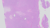
By histology, uterine PEComa may show a circumscribed or infiltrative border, the latter being more common when malignant. They are typically hypercellular but may on occasion be associated with extensive sclerosis (so-called “sclerosing” variant). The majority has a nested growth pattern and is composed of cells having clear to lightly eosinophilic granular cytoplasm. Spindled areas resembling leiomyosarcoma with intersecting fascicles may be seen but the cytoplasm maintains its distinctive granularity. The nuclei can be of varying size and shape and often contain prominent nucleoli. PEComa often demonstrates increased intratumoral vascularity with a staghorn features and tumor may grow in a vasocentric pattern. Cells vary from epithelioid to spindled and have clear to granular eosinophilic cytoplasm. These tumor cells are radially arranged around vessel lumens.
Immunohistochemically, uterine PEComas are characterized by co-expression of smooth muscle and melanocytic markers. In the female genital tract, HMB45 is the most sensitive stain, followed by MiTF and MelanA. Cathepsin K has recently been shown to be consistently and strongly positive in PEComa and is consistently expressed in uterine PEComa. S100 protein is usually negative and helps separate PEComa from melanoma. For smooth muscle markers, desmin is the most sensitive followed by smooth muscle actin and h-caldesmon, which show nearly similar sensitivity. By molecular analysis, some PEComa family tumors lack expression of the tuberous sclerosis- associated TSC2 gene product tuberin.
As only a limited number of gynecologic PEComas have been reported in the English-language literature, criteria for malignancy have yet to be firmly established; nevertheless, based on prior and recent studies, a diagnosis of malignancy can be established if four or more of the following features are present: gross size ≥ 5 cm, significant nuclear atypia, necrosis, lymphovascular invasion, or mitotic index ≥ 1/50 high power fields. Most other PEComas in the uterus should be considered to have uncertain biologic behavior and those with malignant features should be classified as grade 1-3 by French Federation grading criteria (FNCLCC).
The differential diagnosis includes adenosarcoma, endometrial stromal sarcoma (ESS), metastatic melanoma, and, most commonly, leiomyosarcoma. Adenosarcoma, a malignant Mullerian tumor,occurs in a similar yet slightly older age range than PEComa and similarly presents as a polypoid (protruding) mass. However, adenosarcoma usually arises from the endometrium (unless it arises from adenomyosis in the myometrium) rather than myometrium like PEComa. Adenosarcoma is a biphasic neoplasm containing benign glands as the epithelial component and a sarcomatous malignant mesenchymal component. The sarcomatous element often turns out to be sarcoma, NOS (high grade endometrial stromal sarcoma, high grade undifferentiated sarcoma) and/or rhabdomyosarcoma. These sarcomas have a darker low power morphologic appearance than PEComa. Rhabdomyosarcoma additional has epithelioid cells with plump eccentric eosinophilic cytoplasm and immunoreactivity for desmin and skeletal muscle markers, which include myoregulatory proteins nuclear MyoD1 and myogenin (myf4) and is usually negative for SMA. The glands have a leaf-like (phyllodes-like) appearance within sarcomatous stroma. Heterologous elements are present in approximately 10-15% of tumors. Pure rhabdomyosarcoma rarely exists in older females without being part of a mixed mullerian tumor, including adenosarcoma or carcinosarcoma (malignant mixed mullerian tumor). Low grade adenosarcoma may recur in up to 40% of cases, typically in the pelvis or vagina, and distant metastasis has been reported in 5% of cases. High grade sarcomatous differentiation, especially rhabdomyosarcoma, has much worse prognosis. Any rhabdomyosarcomatous component is often treated with extended chemotherapy to cover that phenotype.
Pure ESS is also thought to arise from adenomyosis in the myometrium and has a plexiform staghorn-like vasculature. These tumors are usually low grade, although often infiltrate through myometrium to serosa and in up to a third of cases may have extrauterine extension by plugs of tumor within the vessels of the broad ligament and adnexa. Low grade ESS is typically positive for CD10 and WT1. Smooth muscle phenotype can be present in ESS and when greater than thirty-percent represents hybrid leiomyosarcoma and ESS. Translocation t(7;17) resulting in fusion of two zinc finger genes (JAZF1 and JJAZ1) is present in most low grade endometrial stromal tumors.
Melanoma is exceedingly rare in the uterus and is mostly metastatic. Melanoma should always be considered in a myoid-appearing pleomorphic epithelioid or spindle cell tumor, especially with prominent nucleoli and especially if the nucleoli appear eosinophilic and if dirty necrosis is present. Geographic necrosis is usually “clean” in sarcoma. Melanin pigment may be present and S100 protein would be diffusely positive and desmin and SMA negative.
Leiomyosarcoma is the most common uterine sarcoma and may occur in the setting of patients with multiple leiomyomas, although not thought to arise from a preexisting leiomyoma. These tumors are usually large and soft or gelatinous with necrosis. These are myometrial sarcomas and may undermine endometrium but usually do not present as protruding or polypoid masses in the uterus. Leiomyosarcoma differs by its intensely eosinophilic and less granular cytoplasm, intersecting fascicles of pleomorphic spindled cells, and its strong and diffuse positivity for SMA and desmin and negativity for significant melanocytic marker reactivity. Estrogen receptor is diminished compared with benign leiomyoma yet is usually at least focally present and helps to separate uterine from vascular soft tissue primary, especially with metastases to lung. These are also usually high grade sarcomas (grade 3/3 FNCLCC) and not difficult to separate from benign leiomyoma by their increased cellularity, enlarged pleomorphic cells, increased mitotic activity with atypical forms and geographic necrosis.
Supplementary Questions:
- Which of the following is true regarding uterine malignant PEComa?
- HMB45 is usually negative in these tumors.
- Malignant PEComa is a variant of leiomyosarcoma and is treated the same.
- Patients are usually very young and have other myomelanocytic tumors.
- Patients may present with abnormal uterine bleeding.
- Tumors arise from the endometrium and extend into the uterine cavity.
- What would be a comprehensive immunohistochemical panel for uterine sarcoma?
- Calretinin, CK5/6, pankeratin, D240, MOC31, WT1
- CD34, CD31, D240, Fli-1, Factor VIIIrag, GLUT-1, CD3, CD20, CD79a
- EMA, pankeratin, CK5/6, CK7, CK20, S100 protein, CK18
- S100 protein, HMB45, Melan-A, Mitf, Tyrosinase, pankeratin
- WT1, CD10, ER, SMA, Desmin, HMB45, pankeratin, S100
- All of the following histologic criteria are important to distinguish benign or uncertain from high-grade malignant mesenchymal tumor in the uterus except:
- Atypical mitoses
- Geographic necrosis
- Increased mitoses
- Infiltrative growth pattern
- Polypoid growth pattern
References
- Fadare O. Uterine PEComa: appraisal of a controversial and increasingly reported mesenchymal neoplasm. Int Semin Surg Oncol. 2008;5:7.
- Folpe AL et al. Perivascular epithelioid cell neoplasms of soft tissue and gynecologic origin: a clinicopathologic study of 26 cases and review of the literature. Am J Surg Pathol. 2005;29:1558-1575.
- Italiano A, Delcambre C, Hostein I, et al. Treatment with the mTOR inhibitor temsirolimus in patients with malignant PEComa. Ann Oncol. 2010;21:1135-1137.
- Keylock JB, Fanburg-Smith JC, et al. Renal angiomyolipoma in the first two years of life. A clincopathologic study of 44 cases. Modern Pathology. 2009; 22 suppl 1:175A, abstract 791.
- Schoolmeester JK, Howitt BE, Hirsch MD, Dal Cin P, Quade BJ, Nucci MR. Perivascular epithelioid cell neoplasm (PECome) of the gunecologic tract: clinicopathologic and immunohistochemical characterization of 16 cases. Am J Surg Pathol. 2014;38(2):176-178.
- The European Chromosome 16 Tuberous Sclerosis Consortium. Identification and characterization of the tuberous sclerosis gene on chromosome 16. Cell. 1993;75:1305-1315.
Author
2015
Julie C. Fanburg-Smith, MD
Surgical Pathology Committee
Washington, DC
Answer Key
- Patients may present with abnormal uterine bleeding (d).
- WT1, CD10, ER, SMA, Desmin, HMB45, pankeratin, S100 (e).
- Polypoid growth pattern (e).