- Home
- Member Resources
- Pathology Case Challenge
- Thigh mass
Clinical Summary
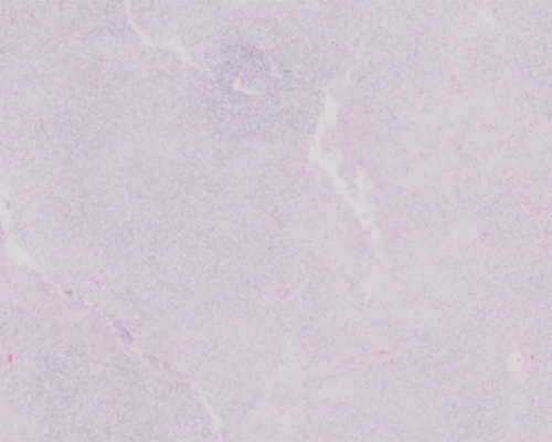
A 47-year-old woman presents with a thigh mass. Upon computed tomography (CT) scan, a 5.2 cm non-homogenously enhancing mass is identified in deep soft tissue. The patient undergoes resection of this thigh mass. Grossly, the specimen is well circumscribed and has a focally disrupted smooth membranous pseudocapsule.
Cut sections show tan-white nodular surfaces with focal gelatinous areas and some hemorrhagic foci.
Master List of Diagnoses:
- Fat atrophy
- Lipoblastoma
- Myxofibrosarcoma
- Myxoid liposarcoma
- Round cell liposarcoma
- Well-differentiated liposarcoma with extensive myxoid changes
Archive Case and Diagnosis
This case first appeared as Performance Improvement Program in Surgical Pathology (PIP) 2016, case 15, and is a myxoid liposarcoma.
Criteria for Diagnosis and Comments
Sections from the tumor show bland fusiform and spindled to ovoid cells in a myxoid matrix with arborizing delicate and plexiform capillary vasculature and immature, primarily univacuolated and bivacuolated, lipoblasts. Pools of extracellular mucinous stroma are observed on some slides. Some sections also show areas of increased cellularity around vessels yet with maintenance of stroma; it is the round cells in the stroma that coalesce back to back without intervening stroma for the criteria of the round cell component. Sometimes the round cell component can have cytoplasmic clear vacuoles. Criteria for round cell liposarcoma includes at least 5% of a round cell component in addition to myxoid areas for classification round cell/myxoid liposarcoma. In addition, when the round cells are increased but not back to back, this feature is called “cellular” myxoid liposarcoma. The presence of a round cell component may increase the grade and portends a worse prognosis than pure myxoid liposarcoma. These are usually graded as grade 2 or grade 3 sarcoma on a 3-tiered system as these tend to respond well to adjuvant therapy, particularly those with more myxoid than round cell change.
Myxoid liposarcoma accounts for about one-third to one-half of all liposarcomas and tends to occur in a younger age group compared with other subtypes of liposarcoma, with a peak incidence in the fifth decade and many patients in their third and fourth decades. It develops preferentially in the lower extremities and buttock and is controversial to occur in the retroperitoneum, with more of those cases representing myxoid change in well- or de-differentiated liposarcoma. Indeed, a myxoid liposarcoma in the retroperitoneum should strongly suggest the possibility of a metastasis. These tumors embrace a continuum of lesions ranging from myxoid tumors with less cellularity to poorly differentiated “round cell” tumors in which lipoblastic differentiation is inconspicuous or appears as cytoplasmic clearing. Histologically, pure myxoid liposarcomas bear a marked similarity to developing fetal fat or atrophic fat. At low power, the classic variant is composed of a paucicellular population of small bland fusiform or round cells in a myxoid matrix composed of hyaluronic acid. A delicate plexiform capillary vascular network provides an important clue for diagnosis. Typically, immature spindle cells intermingle with multivacuolar or unilocular lipoblasts, which are more prominent at the periphery of the lesion. Mitotic figures are typically rare or absent. Frequently pools of stromal mucin impart a pseudolymphangiomatous or pseudoalveolar appearance to the process.
Myxoid and round cell liposarcoma are the same genetic entity. Nearly all myxoid, myxoid/round, and pure round cell liposarcomas are characterized by t(12;16)(q13;p11) translocation. This molecular alteration results in fusion of the DDIT3 (formerly known as CHOP) gene on chromosome 12 with the FUS gene on chromosome 16. The chimeric FUS-DDIT3 gene gives rise to at least three fusion transcripts, one of which (type II) has been identified in most myxoid liposarcomas. The behavior of these tumors is dependent on the amount of round cell differentiation present, which can be measured by a three- or two-tiered system classification employed by Kilpatrick and Antonescu et al, respectively. Age (> 45 years), percent of round cell differentiation (> or = 25%), and the presence of spontaneous tumor necrosis have been shown to be significantly associated with a poor prognosis. It has also been shown that p53 overexpression is a predictor of unfavorable outcome in these tumors. These tumors, even the paucicellular variant, can metastasize to lung, bones, retroperitoneum, and other soft tissue sites, there is an overall 20-40% metastatic potential.
In a large study, it has been determined that myxoid liposarcomas lose their differentiation as they transition from a more cellular lesion (liposarcoma in-transition: 25-50% cells to stroma ratio) to a “round cell” liposarcoma oftentimes composed of primitive cells with high nucleocytoplasmic ratios and round cell transformation (>75% cells to stroma ratio). These tumors acquire a “round cell” appearance in one of two ways: 1) pure round cell nodules composed of primitive round cells without any myxoid stroma, or 2) the gradual progression towards round cells areas with areas of transition. The solid round cell areas can be difficult to identify as liposarcoma, unless an adjacent myxoid region is identified, or back to back monovacuolated lipoblasts are appreciated, or S100 protein is positive. In these situations, differential diagnosis of round cell sarcomas, such as rhabdomyosarcoma, poorly differentiated synovial sarcoma, and Ewing sarcoma/primitive neuroectodermal tumor or lymphoma may be considered and ancillary studies are required to confirm the diagnosis. In these situations, immunohistochemical stains including CD99, leukocyte common antigen (LCA), cytokeratin, myogenin and FLI-1 can be helpful.
Occasionally well-differentiated liposarcomas may have areas of myxoid appearance and exhibit widened septa, heterogeneic low power appearance, and at least focally a pleomorphic atypical lipocytic cells. However, they typically lack the characteristic delicate vasculature seen in myxoid liposarcoma. These tumors also lack the FUS-DDIT3 fusion gene and show amplification of MDM2 and are positive for MDM2 by immunohistochemistry.
Myxofibrosarcoma (previously known as myxoid malignant fibrous histiocytoma) demonstrates a greater degree of nuclear atypia and pleomorphism and possesses ropy coarse vasculature dripping with atypical tumor nuclei and pseudolipoblasts that contain hyaluronic acid rather than lipid.
Fat atrophy can occur in adults and is often due to malnutrition or previous injection site or other. This has adipocytes that are small and appear to be monovacuolated lipoblasts with serous myxoid stromal change and prominence of capillary vessels, so at high power, this can look like a myxoid liposarcoma. Low power will reveal the organized arrangement of the retained fat lobules. This process can occur in the same age group and anatomic distribution of myxoid liposarcoma and is an important differential consideration. Silicone granulomas can also have this myxoid liposarcoma like monovacuolated lipoblast like appearance, but this is positive for CD68 (representing engulfing histiocytes) and negative for S100 protein, as observed in liposarcoma.
Lipoblastoma is a tumor of infancy and early childhood and is composed of irregular small lobules of immature fat cells in different stages of development separated by connective tissue septa of varying thickness and a prominent myxoid matrix. Some of these tumors can show a plexiform vascular pattern, reminiscent of myxoid liposarcoma. However, lipoblastoma is more lobulated than myxoid liposarcoma, occurs in a younger population, and lacks nuclear atypia and areas of increased cellularity. Lipoblastoma is characterized by aberrations in 8q11-13 and lacks the characteristic t(12;16) translocation seen in myxoid liposarcoma. As lipoblastoma matures, it appears as a classic lipoma with retained dense fibrous septa. This would not be in the differential diagnosis for a young adult with myxoid/round cell liposarcoma.
Supplementary Questions
- Which of the following findings is not commonly found in myxoid liposarcoma?
- Delicate plexiform capillary vascular networks are present.
- Mitotic figures are frequently observed.
- Occasionally, pools of mucinous stroma can be seen.
- Paucicellular population of small, bland, fusiform or round cells in a myxoid matrix are present.
- Which of the following statements is true regarding the diagnosis?
- A single FISH test for DDIT3 (DDIT3 was formerly known as CHOP) is not helpful in the diagnosis.
- A translocation involving chromosomes 12 and 16 is identified in a minority of myxoid liposarcoma cases
- A translocation involving chromosomes 12 and 22 resulting in a DDIT3/EWSR1 fusion gene is the most common translocation in the myxoid liposarcomas.
- FUS/DDIT3 fusion gene is the most common translocation observed in myxoid/round cell liposarcoma.
- Which of the following statements is true regarding the prognosis of myxoid liposarcoma?
- Age has no prognostic value.
- Round cell component and presence of necrosis are predictors of unfavorable outcome in patients with myxoid liposarcomas.
- These tumors never metastasize.
- Tumor shows no effect with radiation and chemotherapy.
References
- Fritchie KJ, Goldbum JR, et al. The expanded histologic spectrum of myxoid liposarcoma with an emphasis on newly described patterns: implications for diagnosis on small biopsy specimens. Am J Clin Pathol. 2012;137(2):229-239.
- Kilpatrick SE, Doyon J, Choong PF, Sim FH, Nascimento AG. The clinicopathologic spectrum of myxoid and round cell liposarcoma. A study of 95 cases. Cancer. 1996;77(8):1450-1458.
- Weiss S, Goldblum J. Enzinger & Weiss’s Soft Tissue Tumors. 5th ed. Philadelphia, PA: Mosby; 2008.
Author
2016
Kirtee Raparia, MD
Surgical Pathology Committee
Northwestern University
Chicago, IL
Answer Key
- Mitotic figures are frequently observed. (b)
- FUS/DDIT3 fusion gene is the most common translocation observed in myxoid/round cell liposarcoma. (d)
- Round cell component and presence of necrosis are predictors of unfavorable outcome in patients with myxoid liposarcomas. (b)