Clinical Summary
A 56-year-old man presents with an incidental 5.2 cm gastric tumor (identified on imaging after a motor vehicle accident). Esophagoduodenoscopy shows a submucosal mass lesion in the gastric antrum (mucosal biopsies are normal). Subsequent endoscopic ultrasound (EUS) shows the mass to be centered in the muscularis propria; a needle biopsy is done and is diagnostic. An abdominal and pelvic computed tomography scan with contrast shows no evidence of metastatic disease. After discussion at a multidisciplinary tumor conference, a laparoscopic gastric wedge resection is performed. The tumor is grossly free of the margins and has a solid white cut surface without apparent necrosis. Immunohistochemistry shows labeling in the neoplastic cells for DOG1 and smooth muscle actin (focal), but no labeling for KIT (CD117), S-100, or β-catenin. Representative slides of this resected tumor are presented here.
Master List of Diagnoses
- Gastrointestinal stromal tumor (GIST)
- Inflammatory fibroid polyp
- Inflammatory myofibroblastic tumor
- Intraabdominal fibromatosis (desmoid tumor)
- Leiomyoma
- Leiomyosarcoma
- Schwannoma
Archive Case and Diagnosis
This case first appeared as Performance Improvement Program in Surgical Pathology (PIP) 2016, case 19, and is a gastrointestinal stromal tumor (GIST).
Criteria for Diagnosis and Comments
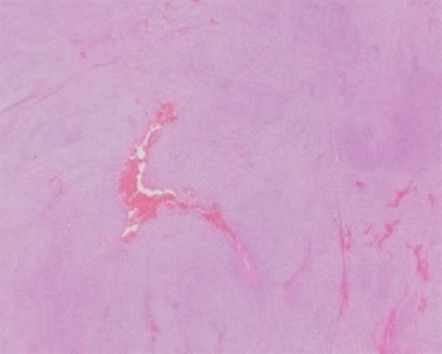
This is a moderately cellular monotonous spindle cell neoplasm with abundant eosinophilic cytoplasm and uniform oval nuclei with dispersed chromatin, no nucleoli and prominent perinuclear vacuoles. There is no nuclear pleomorphism or necrosis. Some sections show areas of stromal myxoid change and hyalinization. The histomorphology in conjunction with the presence of DOG1 (Discovered On GIST 1) immunohistochemical labeling are diagnostic of a gastrointestinal stromal tumor (GIST). By the American Joint Committee on Cancer (AJCC) 7th edition criteria this GIST is low-grade (mitotic figures are <5 mitotic figures per 5 mm2).
Gastrointestinal stromal tumors (GISTs) are uncommon neoplasms that can be seen in patients of any age but are most commonly seen in the middle-aged and elderly. They are most commonly found in the stomach and ileum, rarely in the proximal small bowel, colon, or extraintestinal sites (omentum, mesentery or retroperitoneum), and exceptionally in the appendix and esophagus. GISTs are typically incidental findings either on imaging (as in this case) or an incidental surgical finding. When symptomatic the most common presentations include gastrointestinal (GI) bleeding, abdominal pain, nausea, vomiting, and weight loss.
As in this case, GISTs of the GI tract are typically solitary and centered in the wall. Rarely GISTs can be multifocal; multifocal GISTs are more commonly seen in pediatric patients and in GIST syndromes (familial GIST syndrome, neurofibromatosis type 1, the Carney triad, and Carney-Stratakis syndrome). Cross section typically shows homogenous cut surfaces. Although hemorrhage, necrosis, and cystic change can be seen on occasion, they are not predictors of a higher risk of progressive disease. GISTs have one of three main histologic patterns; (1) pure spindled cell (most GISTs), (2) mixed-spindled and epithelioid, (3) pure epithelioid cells (usually only seen in the stomach).
Immunohistochemistry is useful in the diagnosis of GIST, particularly KIT immunohistochemistry, which is specific marker and is positive in 95% of GISTs. A similarly specific and sensitive marker is DOG1, which is especially helpful in KIT-negative GISTs as the majority of these are positive for DOG1. Other markers that can be seen in GIST, and can confuse the differential diagnosis, include CD34 (70%), smooth muscle actin (30-40%), S-100 and desmin (5%, usually focal), and keratin (1-2%, weak/focal).
The vast majority of GISTs have mutually exclusive mutations in one of two tyrosine kinases genes that lead to constitutive tyrosine kinase activity; 75% in KIT and 10% in PDGFRA. A small minority of GISTs (approximately 8%) have mutations in one of the SDH subunit genes (gene for mitochondrial succinate dehydrogenase complex), these “SDH-deficient GISTs” can be identified by immunohistochemistry (particularly loss of SDHB) and have a particular phenotype; they are often multifocal, have a predilection for young adults and children, commonly have an epithelioid component, metastasize to lymph nodes, and have a indolent course even in the presence of metastatic disease. In contrast, the prognosis of KIT- and PDGFR- mutated GISTs is not associated with the specifics of the mutation. Rare cases have mutations with BRAF or NF1 gene. A small percentage of GISTs have no known mutation, thus mutational analysis is not required to diagnose GIST.
GISTs have variable behavior from benign to overtly malignant. The risk of progressive disease (defined as metastatic disease or tumor-related death) is divided into four categories (none, low, moderate, or high). The best predictors of progressive disease risk in GIST are the anatomic site, tumor size, and mitotic activity; these are the parameters in the published data used by the National Comprehensive Cancer Network (NCCN) task force to assess progressive risk of disease and used in AJCC Staging of GISTs (see table below). Mitotic counts in GIST are the total number of mitotic figures in a 5 mm2 area (this equates to twenty 40x fields in most modern microscopes) and this count solely defines the grade of the GIST (low-grade <5 per 5 mm2 and high-grade >5 per 5 mm2). A single AJCC staging scheme is used for all GI tract GISTs without regard to specific location. Unique to staging of malignancies of the GI tract, GIST staging incorporates the grade into the determination of stage group (eg, depending on grade [mitotic count], pT1 can be either stage IA or II). While the risk of progressive disease increases with increasing AJCC stage, notice that stage-risk correlation is not the same over all sites (eg, stage II is moderate risk in the stomach, but high risk of progressive disease in all other GI sites). When GISTs do metastasize it is usually within the first two years and limited to liver and serosal lining of the abdominal cavity. Nodal metastases and extra-abdominal metastases (lungs, bone, soft tissue and intracranial) are rare. Most gastric GISTs, like the presented case, are indolent lesions with at most low risk of progressive disease.
| Risk Assessment of Primary GIST (with corresponding AJCC staging)* | |||||||
|---|---|---|---|---|---|---|---|
| Tumor Parameters | AJCC | Risk of Progressive Disease (%)** | |||||
| Mitotic Rate (AJCC Grade) | Size (AJCC pT) | N & M status | Stage Group | Gastric | Duodenum | Jejunum/Ileum | Rectum |
| ≤5 per 5 mm2 (low grade) | ≤2 cm (pT1) | N0, M0 | Stage IA | None (0%) | None (0%) | None (0%) | None (0%) |
| >2 - ≤5 cm (pT2) | N0, M0 | Stage IA | Very Low (1.9%) | Low (8.3%) | Low (4.3%) | Low (8.5%) | |
| >5 - ≤10 cm (pT3) | N0, M0 | Stage IB | Low (3.6%) | ## | Moderate (24%) | ## | |
| >10 cm (pT4) | N0, M0 | Stage II | Moderate (10%) | High (34%) | High (52%) | High (57%) | |
| >5 per 5 mm2 (high grade) | ≤2 cm (pT1) | N0, M0 | Stage II | None # | ## | High # | High (54%) |
| >2 - ≤5 cm (pT2) | N0, M0 | Stage II | Moderate (16%) | High (50%) | High (73%) | High (52%) | |
| >5 - ≤10 cm (pT3) | N0, M0 | Stage IIIA | High (55%) | ## | High (85%) | ## | |
| >10 cm (pT4) | N0, M0 | Stage IIIB | High (86%) | ## | High (90%) | High (71%) | |
| Any | Any | N1 or M1 | Stage IV | ||||
* Adapted from table published in CAP GIST protocol using data consolidated in NCCN task force on management of GIST with added data from the AJCC staging manual 7th edition (see references)
** Defined as metastasis or tumor-related death
# Denotes small number of cases
## Insufficient data
Spindled neoplasms that can be seen to primarily involve the stomach also include inflammatory fibroid polyp (IFP), inflammatory myofibroblastic tumors (IMT), and schwannoma. The presence of an inflammatory infiltrate in all three of these entities can be a helpful clue to point away from GIST, which typically has no prominent associated inflammatory component; IFP has a marked eosinophil infiltrate, IMT lymphoplasmacytic inflammation, and schwannoma commonly a lymphoid infiltrate. None of these three entities label for KIT or DOG1 by immunohistochemistry. IFP, like GIST, forms intramural masses and has a cell component that is CD34 positive by immunohistochemistry (the stellate cell); however, these stellate cells are negative for KIT and DOG1. IMT has a distinctive immunohistochemical labeling pattern (labeling for anaplastic lymphoma kinase [ALK]) and, in contrast to most GISTs, it commonly afflicts children and young adults. Schwannoma and GIST can look essentially identical on H&E (including the presence of Verocay bodies) but schwannoma has a distinctive immunohistochemical labeling pattern (S-100 positive in a strong diffuse pattern).
Intraabdominal fibromatosis (desmoid tumor) primarily involves the mesentery or omentum. If there is involvement of the GI tract it is secondary involvement from the outside and with a leading edge of finger-like projections in contrast to the mural centered GIST with a pushing border. In addition, desmoids have collagenous background often with keloid type collagen and demonstrate nuclear labeling for beta-catenin by immunohistochemistry, not seen in GIST, and, in almost all cases, are KIT and DOG1 negative.
By stomach location alone, a smooth muscle neoplasm is unlikely in this case (GI tract leiomyoma, in contrast to GIST, are typically limited to the colorectum and esophagus, and leiomyosarcoma are rarely seen anywhere in the GI tract). The immunohistochemical profile of leiomyomas and leiomyosarcomas provides a clear distinction from GIST, with these smooth muscle neoplasms showing diffuse immunohistochemical labeling for desmin and smooth muscle actin and absence of KIT or DOG1 labeling. Leiomyosarcoma in addition has marked nuclear pleomorphism almost never seen in GIST expect for the exceptionally rare dedifferentiated GIST.
Primary therapy for GIST is surgical with the aim of complete gross total resection and ideally histologically-negative margins. Given the limited intramural extension and low incidence of lymph node metastasis, local regional resection without lymphadenectomy, like the partial gastrectomy in this case, provides adequate surgical treatment. Some GISTs require no immediate treatment. GISTs less than 2 cm may be watched, particularly if no high-risk EUS features are present (eg, no irregular border, cystic spaces, ulceration, echogenic foci, and heterogeneity).
There are three tyrosine kinase inhibitors (TKIs) that have established efficacy in the treatment of GIST, imatinib, sunitinib, and regorafinib; their role depends on the clinical scenario. TKIs are used as a primary therapy in unresectable disease, or theoretically resectable disease in high-risk surgical patients. TKIs are used as adjuvant therapy with GISTs that are found to be moderate or high risk of progressive disease, have gross residual disease (R2 resection) or metastatic disease after resection. The dosage and choice of a particular agent relies in part on the specific mutation (gene and exon), thus the NCCN recommendation is to do mutational analysis in GISTs where TKI therapy is a consideration.
Supplementary Questions
- Which of the following statements regarding AJCC staging of gastrointestinal stromal tumors is true?
- All gastrointestinal stromal tumors of the gastrointestinal tract use the same AJCC staging system without regard to specific location in the gastrointestinal tract.
- Gastrointestinal stromal tumors that involve the serosa are pT4.
- Tumor grade does not affect stage grouping.
- Tumor grade used by the AJCC is based on nuclear pleomorphism.
- When establishing the mitotic count in gastrointestinal stromal tumor, the area to be evaluated equates to the total number of mitotic figures in 50 high power fields of a modern 40x objective.
- In reference to the 5.2 cm low-grade gastric gastrointestinal stromal tumors presented here, which of the following is true?
- The absence of hemorrhage, necrosis, and cystic change on gross evaluation in this case portends a lower risk of progressive disease.
- The NCCN would recommend mutational analysis in this case to further support the diagnosis.
- The solitary nature of this lesion is unusual for a sporadic gastrointestinal stromal tumor.
- The surgery performed here, a local resection without lymphadenectomy, is an appropriate surgery for gastric gastrointestinal stromal tumor.
- This lesion is AJCC stage IB and has low risk of progressive disease and in fact all stage IB gastrointestinal stromal tumors of the gastrointestinal tract have low risk of progressive disease irrespective of anatomical site.
- Regarding the treatment of gastrointestinal stromal tumors, which of the following statements is true?
- The NCCN recommends mutational analysis for all gastrointestinal stromal tumors.
- The NCCN recommends the adjuvant use of tyrosine kinase only with gastrointestinal stromal tumors with a high risk of progressive disease.
- The NCCN recommends the adjuvant use of tyrosine kinase inhibitors after gross total surgical resection in gastrointestinal stromal tumors whose pathology shows microscopically positive margins (R1).
- The principles of surgical resection of gastrointestinal stromal tumors differ from gastrointestinal tract carcinomas as lymphadenectomy is not generally indicated in gastrointestinal stromal tumors due to the low incidence of lymph node metastasis.
- Tyrosine kinase inhibitors are used only as adjuvant therapy.
References
- Demetri GD, von Mehren M, Antonescu CR, et al. NCCN task force report: Update on the management of patients with gastrointestinal stromal tumors. J Natl Compr Canc Netw. 2010;8 Suppl 2:S1-41; quiz S42-4.
- Downs-Kelly E. Chapter 30 - mesenchymal tumors of the gastrointestinal tract. In: Odze R, Goldblum J, eds. Surgical Pathology of the GI Tract, Liver, Biliary Tree and Pancreas. Third ed. Philadelphia, PA: Saunders; 2015:822.
- Hameed M, Corless C, George S, et al. Template for Reporting Results of Biomarker Testing of Specimens from Patients With Gastrointestinal Stromal Tumors. Arch Pathol Lab Med. 2015. [Epub ahead of print] http://www.cap.org/apps/docs/committees/cancer/cancer_protocols/2014/gist-biomarker-14protocol-1000.pdf. Accessed August 15, 2015.
- Miettinen M, Sobin LH, Lasota J. Gastrointestinal stromal tumors of the stomach: A clinicopathologic, immunohistochemical, and molecular genetic study of 1765 cases with long-term follow-up. Am J Surg Pathol. 2005;29(1):52-68.
- Rubin BP, Blanke CD, Demetris AJ, et al. CAP Protocol for the Examination of Specimens from Patients with Gastrointestinal Stromal Tumor (GIST). http://www.cap.org/apps/docs/committees/cancer/cancer_protocols/2014/gist-14protocol-3100.pdf. Accessed August 15, 2015.
- Rubin BP, Heinrich MC. Genotyping and immunohistochemistry of gastrointestinal stromal tumors: An update. Semin Diagn Pathol. 2015. DOI: https://dx.doi.org/10.1053/j.semdp.2015.02.017. Accessed August 15, 2015.
- von Mehren M, Randall RL, Benjamin RS, Boles S. NCCN Clinical Practice Guidelines in Oncology Soft Tissue Sarcoma (NCCN guidelines®). http://www.nccn.org/professionals/physician_gls/pdf/sarcoma.pdf. Accessed August 15, 2015.
Author
2016
H. Parry Dilworth, MD
CAP Surgical Pathology Committee
Hospital Pathology Associates
Minneapolis, MN
Answer Key
- All gastrointestinal stromal tumors of the gastrointestinal tract use the same AJCC staging system without regard to specific location in the gastrointestinal tract. (a)
- The surgery performed here, a local resection without lymphadenectomy, is an appropriate surgery for gastric gastrointestinal stromal tumor. (d)
- The principles of surgical resection of gastrointestinal stromal tumors differ from gastrointestinal tract carcinomas as lymphadenectomy is not generally indicated in gastrointestinal stromal tumors due to the low incidence of lymph node metastasis. (d)