- Home
- Member Resources
- Pathology Case Challenge
- Spleen copy
Clinical Summary
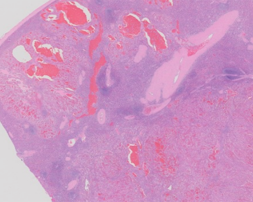
A 56-year-old woman presents with significant weight loss. She was in her usual state of health until five months ago, when she noted a mass over her left lower thorax. A magnetic resonance imaging (MRI) scan of the back is performed, which reveals a lipoma of the left lower thorax. The MRI incidentally notes splenomegaly with nodularity and hypodensities within the spleen suspicious for lymphoma. Physical examination confirms splenomegaly 5.0 cm below the left costal margin. Splenectomy is performed, and the 540-gram spleen measures 15.0 cm x 13.0 cm x 7.0 cm. The capsule is intact, and the cut splenic surface shows multiple dark red-brown spongy vascular nodules. The H&E section represents one of the nodules.
Master List of Diagnoses
- Angiosarcoma
- Hamartoma
- Hemangioma
- Kaposi sarcoma
- Littoral cell angioma
- Lymphangioma
- Sclerosing angiomatoid nodular transformation of spleen
Archive Case and Diagnosis
This case first appeared as Performance Improvement Program in Surgical Pathology (PIP) 2016, case 35, and is a littoral cell angioma.
Criteria for Diagnosis and Comments
This case demonstrates littoral cell angioma. The histological sections consist of splenic architecture effaced by multiple irregular cystic lesions with vascular proliferation lined by distinctive plump cells. The plump cells “fall off” of the endothelium and are seen sitting within the vascular lumens, along with histiocytes. In rare areas the plump cells form vaguely papillary structures. The lining cells are tall endothelial cells, protruding into the lumen, with little atypia. There is little to no normal white pulp or red pulp in some of the sections examined, except on slides where focal white pulp with germinal centers is found at the periphery near the capsule. The lesional cells are positive for CD31, CD68, and CD163.
This is a relatively rare vascular neoplasm of the spleen commonly found in association with other neoplasms; it is thought to arise from the red pulp sinus lining cells known as littoral cells. This lesion is unique to the spleen as there is no counterpart in other tissues, but it can also occur within accessory spleen. Littoral cell angioma may be seen in all age groups with a median age of 55 and a wide age range (5–95 years) and is equally found in males and females.
Most of the patients are asymptomatic, and the lesion is most commonly found incidentally during imaging studies (as it was in this case) or after splenectomy. Abdominal pain is the most common presenting symptom and laboratory tests are generally unremarkable, other than sometimes showing anemia or thrombocytopenia.
Littoral cell angioma is benign, and splenectomy without adjuvant therapy is curative. These tumors are generally multiple, non-encapsulated, nodular masses, and only rarely are the lesions solitary. There is a spongy appearance that may be partially cystic on gross inspection. They can enlarge the spleen and rarely cause splenic rupture. The lesions range in size from 1.0 to 20.0 cm with unremarkable surrounding splenic tissue. The spleen usually has normal weight (< 200 grams) in ~ 33% of cases. The lesion consists of multiple red-brown nodules representing the vascular cystic areas.
Histologically, there are open vascular spaces (cystic areas) that are lined by distinctive plump round to cuboidal epithelial cells (littoral cells). The epithelial cells protrude into the vascular lumens. Rarely, papillary projections of these cells can extend/fall off into the vascular lumen. The lining cells are medium in size with bland, round to slightly irregular lumen with dispersed chromatin. There is also ample pink cytoplasm. Normal splenic sinuses have cells that are more flattened as compared to the cells in littoral cell angioma. Mitotic figures are indistinct. The splenic capsule typically remains intact, except in the rare cases that have caused splenic rupture. Small amounts of inflammatory cells (neutrophils, histiocytes, small lymphocytes) may be associated with the vasculature, but is usually not a distinct component of the lesion. The splenic parenchyma surrounding the littoral cell angioma usually consists of white pulp and red pulp that is unremarkable.
Littoral cell angiomas are uniquely positive for endothelial and vascular markers, (Factor VIII, CD31, ERG, CD68, and CD163), although negative for CD34. They also do not typically express CD8, which normal splenic littoral cells do. A recent study determined that they are negative for WT1, differentiating littoral cell angioma from other splenic vascular lesions, which are all positive for WT1.
Included in the differential diagnosis are other benign vascular lesions (sclerosing angiomatoid nodular transformation, hemangiomas, and lymphangioma), Kaposi sarcoma, angiosarcoma, and hamartoma. Both histology and immunohistochemical staining can be helpful in making the diagnosis.
Sclerosing angiomatoid nodular transformation (SANT) is a rare, benign vascular lesion of unknown etiology, which is composed of multiple confluent vascular nodules with a unique composition of three variant vessels, including capillaries, venules, and sinusoids, with prominent intervening bands of fibrosis. Immunohistochemically, the capillaries are CD34, CD31, ERG, and WT1 positive, but are CD8 negative. Sinusoids are CD31+ and CD8+ and CD34 negative. The small veins are CD31 positive, but negative for CD8 and CD34. Myofibroblasts in the fibrous tissue are positive for smooth muscle actin. SANT would not contain the littoral cell-lined cysts of a littoral cell angioma and is usually a unifocal lesion unlike littoral cell angioma. Littoral cell angioma does not show the three variant vessels or the fibrosis of SANT.
Hemangiomas are the most common primary tumors of the spleen, which are a benign vascular proliferation. Grossly, they are usually bloody and cystic. Splenic hemangioma may be cavernous or capillary depending on the size of the vascular spaces, but cavernous types are more common in the spleen. They consist of thin-walled vascular elements without atypia and resemble hemangiomas at other sites. The endothelial cells are positive for vascular markers (Factor VIII and CD31) but lack CD8 expression. In contrast to littoral cell angioma, hemangiomas do not typically show plump cells that fall off into the vascular lumens, and do not express histiocytic markers such as CD68 and CD163.
Lymphangioma is a benign vascular tumor composed of lymphatic vessels. Sections show thin-walled cysts containing clear gross fluid. The lumina lack red blood cells, and instead may be filled with proteinaceous eosinophilic material. Lymphatic endothelial cells are typically flat, unlike littoral cells, and are immunoreactive with D2-40, variably with CD31, CD34, and factor VIII. The flat endothelial lining and presence of D2-40 would distinguish lymphangioma from littoral cell angioma.
Angiosarcoma is a malignant vascular neoplasm made of hemorrhagic nodules with multiple interanastomosing vascular channels, necrosis, and increased mitoses including atypical mitotic figures. Angiosarcoma is immunoreactive with endothelial markers (ERG, CD34 and CD31), but negative for CD8. A littoral cell angiosarcoma is very rare but has been described. It is cytologically and immunophenotypically similar to a littoral cell angioma but has lining cells that are more atypical than seen in a littoral cell angioma. Mitoses and necrosis seen in angiosarcoma would not be seen in littoral cell angioma.
Kaposi sarcoma may also involve the spleen. It is associated with HIV positivity as well as human herpes virus 8. The tumor is composed of spindled flat malignant endothelial cells, often with eosinophilic globules. Littoral cell angioma can be distinguished from Kaposi sarcoma by lacking HHV8 and expressing histiocytic markers.
Hamartomas are rare, well-circumscribed lesions, which are made up of red pulp elements. They are composed of a proliferation of sinuses, similar to that of the normal splenic red pulp. White pulp elements are distinctly not present in these lesions. Hamartomas do not show the plump endothelial lining cells of littoral cell angioma, CD8 is characteristically positive in hamartoma and negative in littoral cell angioma. Vascular immunohistochemical markers should be negative, except in incidental vessels.
Supplementary Questions
- Which of the following is true regarding the typical staining pattern of littoral cell angioma?
- Stains positively with CD31, CD68, and ERG
- Stains positively with CD31, CD68, and WT1
- Stains positively with CD34, CD68, and human herpes virus 8
- Stains positively with CD8, CD163, and ERG
- Stains positively with CD8, CD68, and CD163
- Which of the following is true regarding littoral cell angioma?
- Almost always presents with cytopenias
- Frequently transforms into angiosarcoma
- Occurs exclusively in men
- Occurs exclusively in the elderly
- Only occurs in the spleen
- Which of the following favors a diagnosis of littoral cell angioma, as compared to sclerosing angiomatoid nodular transformation of spleen?
- Bands of fibrosis
- Mitotic figures and necrosis
- Multifocal lesion
- Positive WT1 immunohistochemical staining
- Three variant vessels
References
- Auerbach A. Diagnostic Pathology: Spleen. 1st ed. Salt Lake City, UT: Amirsys Inc. 2014:3:14-15.
- Kaza RK, Azar S, Al-Hawary MM, Francis IR. Primary and secondary neoplasms of the spleen. Cancer Imaging. 2010;10:173-182.
- Leung VA, Tang S, Mahe E, Patlas MN. Littoral cell angioma: Diagnosis by image-guided biopsy. Ann Clin Lab Sci. 2012;42:417-421.
- O’Malley DP, Kim YS, Weiss LM. Distinctive immunohistochemical staining in littoral cell angioma using ERG and WT-1. Ann Diagn Pathol. 2015;19(3):143-145.
- Matuszczak E, Reszec J, Dębek W, Hermanowicz A, Chyczewski L. Is littoral cell angioma of the spleen as rare as previously believed in the pediatric population? Folia Histochem Cytobiol. 2012;50:480-485.
- Venkatanarasimha N, Hall S, Suresh P, Williams MP. Littoral cell angioma in a splenunculus: a case report. Br J Radiol 2011;84(997):e11-31.
Authors
2016
Rebecca Johnson, MD
Pathology Resident
Walter Reed National Military Medical Center
Bethesda, MD
Aaron Auerbach, MD, MPH
Surgical Pathology Committee
The Joint Pathology Center
Silver Spring, MD
Answer Key
- Stains positively with CD31, CD68, and ERG (a)
- Only occurs in the spleen (e)
- Mulitfocal lesion (c)