Clinical Summary
A 66-year-old man presents to his primary care physician with complaints of abdominal discomfort, fullness, nausea and vomiting, especially, after a meal. Physical examination reveals non-tender splenomegaly, which is 7.0 cm below the left costal margin. Complete blood count is normal. A CT scan reveals an enlarged spleen with multiple lesions interspersed within normal appearing parenchyma. A bone marrow biopsy, performed as part of the work-up to exclude a systemic lymphoma, was normal. Upper endoscopy reveals a gastric antral nodule, which on biopsy shows mild chronic gastritis and focal intestinal metaplasia. Staining for H. pylori is negative.
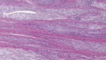
Due to the symptoms, a splenectomy is performed. The 178 g spleen has an intact, nodular gray-tan capsule and measures 10.2 x 7.7 x 4.5 cm. It shows small bluish nodules on the surface and interspersed within the splenic parenchyma on cut sections. No necrosis is seen. An H&E slide of the spleen is given for review.
Tissue is submitted for flow cytometry and does not reveal any abnormalities (ie, a polytypic B-cell population, and unremarkable T-cell population are identified).
Master List
- Hairy cell leukemia
- Littoral cell angioma
- Sclerosing angiomatoid nodular transformation
- Splenic angiosarcoma
- Splenic hemangioma
- Splenic lymphangioma
Archive Case and Diagnosis
This case first appeared as Performance Improvement Program in Surgical Pathology (PIP) 2014, case 17 and is a littoral cell angioma.
Criteria for Diagnosis and Comments
Histologic sections show overall preservation of splenic parenchyma. Within the parenchyma, there are focal areas of vascular proliferation that correspond to the bluish areas present on gross examination. The vascular spaces are characterized by conspicuous lining cells that are tall and foamy (littoral cells) in contrast to normal endothelial cells seen elsewhere which are flat without discernible cytoplasm. The lining cells are positive for CD68 and S100 protein, but negative for Factor VIII and CD34.
This histologic profile along with the immunohistochemical phenotype is diagnostic of littoral cell angioma (LCA).
LCA is a benign distinctive entity of the spleen, composed of splenic sinus lining cells. There are no descriptions of this cell type or littoral cell proliferations in other organ systems in the human body. There is a wide age range with a mean age of 49 years. Splenomegaly, when present, is not prominent. Grossly, LCAs appear mostly as multiple blood filled spongy nodules; occasional spleens may display a solitary (or a dominant) nodule. Histologically, they are characterized by an anastomosing network of vascular channels, which have a pseudo-papillary appearance in crowded areas. Markedly dilated cavernous spaces (vascular lakes) may be seen occasionally. The pathognomonic feature is the littoral cells lining the vascular spaces. The littoral cells are tall cells with regularly indented nuclei, sometimes with a ‘hobnail’ appearance. When dilated vascular spaces are seen, exfoliated littoral cells are seen within the lumina sometimes.
LCA, especially in crowded areas and with high proliferation (ie, multiple mitoses), may be mistaken for angiosarcoma of the spleen. Angiosarcoma of the spleen, similar to angiosarcoma in soft tissues and elsewhere, is characterized by nuclear pleomorphism, high degree of atypia and increased mitosis especially in poorly differentiated lesions. On the other hand, the tall littoral cells in LCA usually have a well-differentiated cytology.
Sclerosing angiomatoid nodular transformation (SANT) is a recently described entity of the spleen. Nodular lesions have varying degrees of fibrosclerotic bands effacing the splenic architecture and a characteristic fibroblastic proliferation inducing sclerosis around vascular channels, some with thrombosis. Extravasated RBCs are frequently seen in SANT, but may also be seen in other vascular lesions of the spleen.
Splenic involvement by hairy cell leukemia can show irregular blood lakes (‘pseudo-sinuses’) that may mimic littoral cell angioma. The blood lakes lack lining cells in hairy cell leukemia.
Lymphangioma and hemangioma of the spleen are focal proliferations of lymphatics and vascular spaces of the spleen. In lymphangioma, the lumina lack cellular elements (i.e. no red cells or white blood cells) and instead, only proteinaceous material is found in the lumina. The endothelial lining is typically flat. In contrast, hemangioma usually contains formed blood elements such as red blood cells and white blood cells. Splenic hemangioma, as can occur in other sites, may be cavernous (more common) or capillary, depending on the size of the luminal spaces. They are lined by flat endothelial spaces and separated by fibrous septa.
Cytomorphology and histopathology of good quality sections usually are enough to diagnose the various entities. Immunohistochemistry may be an ancillary aid in challenging cases. The lining cells of most of the above entities are CD31 (PECAM – platelet endothelial cell adhesion molecule) positive. However, other vascular markers such as CD34 and factor VIII related antigens are typically negative in LCA. In addition, LCA is positive for CD68 and CD21. CD8 expression, which is typically seen in splenic endothelial cells, is typically absent in LCA. Thus, the typical vascular profile of LCA is CD34-/CD68+/CD21+/CD8-; Contrasting to this immuno-phenotype, other hamartomatous lesions and hemangiomas are CD34+/CD68-/CD21-/CD8+.
In summary, LCA is a distinctive entity of the spleen and is characterized by localized proliferation of littoral cells. Clinical prognostic outcome after splenectomy is excellent.
Supplementary Questions:
- Which lesion is composed of a proliferation of endothelial cells forming lumens with formed red blood cells and endothelial-lining cells that express CD34, Factor VIII, and CD8?
- Hairy cell leukemia
- Littoral cell angioma
- Sclerosing angiomatoid nodular transformation
- Splenic lymphangioma
- Splenic hemangioma
- Which entity shows distinctive nodules of radiating fibrosclerotic bands intermingled with occasional medium caliber thrombosed blood vessels?
- Littoral cell angioma
- Sclerosing angiomatoid nodular transformation
- Splenic angiosarcoma
- Splenic lymphangioma
- Splenic hemangioma
- Which of the above entities has the following immunophenotype: CD34-/CD68+/CD21+/CD8-?
- Hairy cell leukemia
- Littoral cell angioma
- Splenic angiosarcoma
- Splenic hemangioma
- Splenic lymphangioma
References
- Arber DA, Strickler JG, Chen YY, Weiss LM. Splenic vascular tumors: a histologic, immunophenotypic, and virologic study. Am J Surg Pathol. 1997;21(7):827-835.
- Fanta PT, Saven A. Hairy cell leukemia. Cancer Treat Res. 2008;142:193-209.
- Fletcher CDM, Bridge JA, Hogendorn P, Mertens F, eds. WHO Classification of Tumours of Soft Tissue and Bone. 4th ed. Lyon, FR: IARC; 2013.
- Warnke RA, Weiss LM, Chan JKC, Cleary ML, eds. Tumors of the lymph nodes and spleen: Atlas of Tumor Pathology. 3rd series Fascicle 14. Washington, DC; AFIP Press; 1995:495-508.
Author
2014
Rajan Dewar, MD, PhD FCAP
Surgical Pathology Committee
Staff Hematopathologist
Department of Pathology
Beth Israel Deaconess Medical Center and Harvard Medical School
Boston, MA
Answer Key
- Splenic hemangioma (e).
- Sclerosing angiomatoid nodular transformation (b).
- Littoral cell angioma (b).