Clinical Summary
A 63-year-old man presents to the emergency room with a ruptured spleen and an emergency splenectomy was done. He carries a diagnosis of a myeloproliferative neoplasm, for the past 2 years and has progressively become pancytopenic. His peripheral blood smear shows a leuko-erythroblastic picture.
Master List
- Classical Hodgkin lymphoma
- Gaucher disease
- Hairy cell leukemia
- Splenic extramedullary hematopoiesis secondary to primary myelofibrosis
- Splenic marginal zone lymphoma
Archive Case and Diagnosis
This case first appeared as Performance Improvement Program in Surgical Pathology (PIP) 2013, case 05, and is splenic extramedullary hematopoiesis secondary to primary myelofibrosis.
Criteria for Diagnosis and Comments
The flow cytometry profile reveals mostly mature polyclonal B-cells, normal T-cells and a population of maturing granulocytes and precursors. A distinct blast population is not seen. Peripheral blood sent for JAK2 V617F mutation is negative, but is positive for MPL W515L mutation.
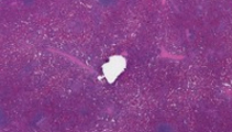
Microscopic sections show marked expansion of the red pulp and intact white pulp. Most of the red pulp is composed of erythroid islands, myeloid precursor cells and megakaryocytes. A subset of megakaryocytes is atypical with "cloudy" condensed chromatin and a few are seen within sinusoidal spaces. A few mitotic figures are noted. The erythroid islands show normoblastic maturation. The myeloid cells are left shifted, but blasts are not increased (CD34 and CD117 stains highlight about 5% of myeloid cells). The white pulp is unremarkable. Reed-Sternberg or Hodgkin cells are not seen. Histiocytes/macrophages are not increased. In a patient with a history of myeloproliferative neoplasm, the findings are consistent with splenic extramedullary hematopoiesis.
Splenomegaly can be seen in many cases of myeloproliferative neoplasm—including chronic myelogeneous leukemia, primary myelofibrosis, polycythemia vera and essential thrombocytosis. In particular, they can be seen in both the cellular phase of the disease, when the neoplastic clonal expansion can cause the splenomegaly. More commonly, the splenomegaly is apparent in myelofibrosis or bone marrow failure, when the spleen becomes the primary organ of hematopoiesis. This patient had progressive cytopenia. The leukoerythroblastic picture seen in peripheral blood was suggestive of myelofibrosis. In addition, DNA studies showed that the patient carried MPL W515L mutation and was negative for JAK2 V617F mutation. Recent studies show that JAK2 V617F is seen in 95% of cases with polycythemia vera and post-polycythemic myelofibrosis. JAK2 V617F is seen only in 50% of patients with primary myelofibrosis. 5-7% of patients with primary myelofibrosis carry a mutation within the MPL W515L/K gene. Many of the morphological changes characteristic of primary myelofibrosis typically seen in the bone marrow are also seen in the spleen. Thus the characteristic megakaryocytic nucleus with condensed cloud like nucleus (as opposed to large megakaryocytes with branched nuclear lobes seen in essential thrombocytosis) is seen in the spleen of this patient. Another feature typical of primary myelofibrosis—intravascular hematopoiesis—seen in the marrow can be seen within the spleen.
Splenic involvement in classical Hodgkin lymphoma is quite common. Gross examination can show nodular lesions. Identifying neoplastic Reed-Sternberg cells or Hodgkin cells is essential for the diagnosis. Rarely, tangentially sectioned megakaryocytes may appear to be of similar size and morphology as Hodgkin/Reed-Sternberg cells. When in doubt, immunostains can help distinguish between the two. Hodgkin cells are CD45 negative, CD30 and CD15 positive. Megakaryocytes are CD42 and CD31 positive.
Gaucher disease is the most common lysosomal storage disease, due to the hereditary deficiency of glucosylceramidase. This disorder also causes pancytopenia and splenomegaly, but the age of onset is typically younger. The spleen is infiltrated by macrophages with engorged cytoplasm, which has a 'crinkled tissue paper' appearance—due to accumulation of glucocerebroside. The infiltration is typically diffuse within the red-pulp. Trilineal cells are not seen. Definitive diagnosis of this disease is by genetic testing.
Splenic marginal zone lymphoma show extensive white pulp involvement and usually grossly visible nodular expansion. Flow cytometry shows B-cells that express CD19, CD20, and are negative for CD5 and CD10 with surface light chain restriction.
Hairy cell leukemia is a differential diagnosis of splenic red pulp expansion. However, hairy cell leukemia causes red-pulp expansion with medium sized lymphocytes, as opposed to extramedullary hematopoiesis which shows mostly trilineage (erythroid, myeloid and megakaryocytic) hematopoiesis. Hairy cell leukemia typically shows a dense and diffuse interstitial/parenchymal involvement. In addition, hairy cell leukemia has a specific flow cytometry immunophenotype (lymphocytes expressing CD19, CD25, CD11c and CD103 with surface light chain restriction).
Splenectomy is not a classical therapeutic or diagnostic intervention in primary myelofibrosis. In a study of 223 patients from the Mayo clinic, survivors of splenectomy (9% operative mortality) had a median survival time of about 27 months. Following splenectomy, patients experienced hepatomegaly and thrombocytosis. About 16.3% of patients also had blast transformation. Splenectomy is considered only as a palliative measure or as a result of traumatic laceration as seen in this patient.
In summary, splenomegaly in myeloproliferative neoplasms can show extramedullary hematopoiesis. Histopathological and ancillary examination should be performed to exclude transformation to acute leukemia and to rule out other disorders.
Supplementary Questions:
- Which of the following entities is characterized by splenic white pulp involvement and by flow cytometry shows a population that is positive for the markers CD19 and CD20 with light chain restriction?
- Classical Hodgkin lymphoma
- Primary myelofibrosis
- Splenic extramedullary hematopoiesis secondary to primary myelofibrosis
- Splenic marginal zone lymphoma
- A patient with HIV infection shows symptoms suggestive of bone marrow failure and pancytopenia. Large binucleate cells with prominent eosinophilic nucleoli are seen in the bone marrow biopsy and within spleen. The large atypical cells are CD30 positive and CD15 positive. What is the diagnosis?
- Classical Hodgkin lymphoma
- Hairy cell leukemia
- Primary myelofibrosis
- Splenic marginal zone lymphoma
- A characteristic morphology seen in primary myelofibrosis which is less common in essential thrombocytosis or chronic myeloid leukemia is the following:
- Enlarged megakaryocytes with branched nuclear lobes
- Megakaryocytes showing compact 'cloud' like nucleus with condensed chromatin
- Megakaryocytes with 'crinkled' tissue paper cytoplasm
- Mononuclear megakaryocytes in tight clusters
References
- Cervantes F. Modern management of myelofibrosis. British Journal of Haematology. 2005;128:583-592.
- Grabowski GA. Phenotype, diagnosis and treatment of Gaucher's disease. Lancet. 2008;372:1263-1271.
- Swerdlow SH, Campo E, Harris NL, et al., eds. World Health Organization Classification of Tumours of Haematopoietic and Lymphoid Tissues. Lyon, France: IARC Press; 2008.
- Tefferi A, Mesa RA, Nagorney DM, Schroeder G, Silverstein MN. Splenectomy in myelofibrosis with myeloid metaplasia: a single institution experience with 223 patients. Blood. 95:2000;2226-2233.
Author
2012
Rajan Dewar, MD, PhD, FCAP
Surgical Pathology Committee
Beth Israel Deaconess Hospital and
Harvard Medical School
Boston, MA
Answer Key
- Splenic marginal zone lymphoma (d).
- Classical Hodgkin lymphoma (a).
- Megakaryocytes showing compact 'cloud' like nucleus with condensed chromatin (b).