- Home
- Member Resources
- Pathology Case Challenge
- Right Ovary
Clinical Summary
A 9-year-old girl presents with a three-week history of lower abdominal discomfort. Upon physical examination a right adnexal mass is palpated, subsequently confirmed by radiologic evaluation. There is no evidence of abnormal secondary sexual characteristics. Surgical resection of the right adnexal structures reveals an intact cystic ovarian tumor with a smooth outer surface, measuring 12.0 cm in greatest dimension. Upon sectioning, close to 30% of the cut surface is cystic and 70% solid. The cystic, smooth-walled, tan areas are filled with serosanguinous fluid; the largest cyst measures 4.5 cm in greatest dimension. The solid component is tan and soft, without gross evidence of necrosis or hemorrhage. A grossly unremarkable fallopian tube is also present. No evidence of metastatic disease or ascites is noted.
Master List
- Adult granulosa cell tumor
- Clear cell, undifferentiated, and transitional cell carcinoma
- Embryonal carcinoma
- Juvenile granulosa cell tumor
- Metastatic malignant melanoma
- Pregnancy luteoma
- Small cell carcinoma, hypercalcemic type
- Steroid cell tumor, not otherwise specified (NOS)
- Thecoma
- Yolk sac tumor
Archive Case and Diagnosis
This case first appeared as Performance Improvement Program in Surgical Pathology (PIP) 2014, case 23 and is a juvenile granulosa cell tumor.
Criteria for Diagnosis and Comments
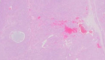
The histologic sections reveal a neoplastic proliferation composed of rounded cells with abundant eosinophilic cytoplasm and hyperchromatic round to oval nuclei; nuclear grooves are rare. Variable mitotic activity is present. There are follicles of varying sizes, some of them filled with basophilic secretions. Neoplastic cells with vacuolated cytoplasm line some follicles and are also noted in scattered foci. Most of the neoplastic cells are arranged in a diffuse pattern alternating with a nodular pattern. In some areas single neoplastic cells and small clusters of neoplastic cells are noted in a mostly edematous connective tissue background. A minor thecomatous component is noted. Call-Exner bodies are not identified. The tumor cells are diffusely immunopositive for inhibin. The microscopic findings are consistent with a juvenile granulosa cell tumor (JGCT).
The category of ovarian sex cord-stromal tumors comprises neoplasms composed of sex cord derivatives (granulosa cells, Sertoli cells) and stromal derivatives (theca cells, Leydig cells, fibroblasts of stromal origin); they account for close to 8% of all ovarian tumors. These neoplasms can occur in pure forms or in various combinations and degrees of differentiation. Tumors of ovarian cell types are commonly estrogenic (only occasionally androgenic), while tumors of testicular cell types are commonly androgenic (only occasionally estrogenic). However, not all ovarian sex cord-stromal tumors are hormonally active.
Granulosa cell tumors (GCTs) are tumors composed of at least a 10% population of neoplastic granulosa cells and account for close to 1.5% of all ovarian tumors.
Two distinct histopathologic subtypes of GCTs are recognized: the adult type and the juvenile type. The adult granulosa cell tumor (AGCT) is the most common subtype (accounting for close to 95% of all GCTs) and mainly occurs in middle aged to postmenopausal women, while the JGCT subtype occurs mostly in children and women <30 years of age. Although uncommon, AGCTs can occur in children and young women and JGCTs can occur in middle age and postmenopausal women.
When JGCTs occur before puberty, close to 80% of the patients develop signs and symptoms of isosexual pseudoprecocity, the result of hormonal tumor activity that may be estrogenic or (rarely) androgenic. In post-pubertal females other signs and symptoms of estrogenic or androgenic hormonal activity may develop, including menstrual irregularities. Clinical presentation commonly includes a palpable adnexal mass. Less common clinical findings include tumor rupture with development of hemoperitoneum, the presence of ascites, and extraovarian tumor spread. An association with Ollier's disease (enchondromatosis) and Maffucci syndrome (enchondromatosis and hemangiomatosis) is noted in the literature.
JGCTs are unilateral in close to 98% of cases, and range in size from 3 to 32 cm in greatest dimension (average diameter is 12.5 cm); they resemble AGCTs grossly. The tumors can be solid or solid and cystic with a smooth or lobulated outer surface. The cysts are mainly smooth lined and can be filled with hemorrhagic and/or clear fluid; rare multicystic and unilocular cystic tumors may be seen. The solid component is soft to firm, and can be tan, gray, yellow, or variegated. Areas of hemorrhage and/or necrosis may be prominent.
Microscopic evaluation usually reveals a solid proliferation of neoplastic granulosa cells associated with a minor follicular component. It is uncommon to identify a uniformly solid (often nodular) cellular pattern or a predominantly follicular pattern. Although rare, a pseudopapillary pattern can also be observed. The follicles of the JGCT can vary in size and shape, but tend to be round to oval and filled with eosinophilic to basophilic fluid (that may be mucicarminophilic in close to 2/3 of cases). Lining the follicles are granulosa cells (that may be arranged in layers) occasionally surrounded by theca cells. On rare occasions, granulosa cells with hobnail contours line the follicles. Call-Exner bodies are rarely present. A variable component of theca cells may be present, however, rare tumors may display areas with a predominant thecomatous/fibrothecomatous component, including the presence of hyaline plaques. If necessary, reticulin stains can assist in differentiating between neoplastic granulosa and theca cells (fibrils surround the individual theca cells, but only surround groups of granulosa cells). Areas of sclerosis and calcification are uncommon. In JGCTs, the granulosa cells commonly exhibit abundant eosinophilic cytoplasm and generally rounded hyperchromatic nuclei with rare nuclear grooves (although some foci of more typical AGCT type cells can be rarely identified). Nuclear atypia varies from minimal to severe (severe atypia is focally present in close to 15% of cases). Mitotic activity is variable and may exhibit abnormal forms, but it is usually higher than that of AGCTs. Cytoplasmic vacuolization characteristic of luteinization can be present in the neoplastic granulosa and/or theca cells (fat stains [e.g., oil red O] can confirm the presence of cytoplasmic lipid). The term "anaplastic JGCT" is used by some experts to designate neoplasms with a sheet-like growth of markedly atypical cells resembling undifferentiated carcinoma that, after extensive sampling, reveals features more characteristic of JGCTs (including the presence of typical follicles). They appear to be highly malignant tumors. The neoplastic granulosa cells can be immunopositive for alpha-inhibin, calretinin, vimentin, S-100 protein, smooth muscle actin, CD10, CD99, Mullerian inhibiting substance, SF-1, CD56, ER, PR, WT-1, AE1/AE3 and CAM 5.2, and immunonegative for SALL4, CK7, and EMA. A missense point mutation in FOXL2 is present in >90% AGCTs, but is rarely present in JGCTs. Trisomy 12 has been demonstrated in AGCTs and JGCTs.
Diagnosis and Comments
Several ovarian lesions are in the differential diagnosis of a JGCT. Here are some important features associated with these lesions that should assist in the evaluation of this type of tumor:
- Some histological features can assist in differentiating between Adult granulosa cell tumors and JGCTs. The neoplastic cells of the AGCTs usually have scant cytoplasm and uniform, angular to oval, often grooved ("coffee bean") nuclei with prominent nucleoli. Mitotic activity is variable, but not as prominent as that observed in JGCTs. The follicles of the AGCTs are more regular in size and shape than the follicles of the JGCTs. In AGCTs, Call-Exner bodies are prominent and luteinization is rare (except when the patients are pregnant). These tumors also display a variety of architectural patterns (frequently admixed), including trabecular and corded, insular, and gyriform patterns. GCTs of both subtypes have similar immunohistochemical profiles.
- The ovarian small cell carcinoma of the hypercalcemic type (SCC HCT) also occurs in children and young women. The neoplastic cells of the SCC HCT are small, have scanty cytoplasm, and are usually arranged in sheets and cords; mitotic activity is very brisk. Tumor necrosis is not uncommon and they commonly exhibit follicle-like structures with eosinophilic (or, rarely, basophilic) luminal fluid. SCC HCT are not estrogenic, commonly present in association with hypercalcemia, and are very aggressive, presenting (in close to 50% of cases) with tumor dissemination beyond the ovary. Mucinous epithelium can be seen in close to 12% of cases, but they lack theca cells. Immunostaining is variable. They can be immunopositive for EMA, vimentin, CAM 5.2, NSE, and chromogranin but are, in general, immmunonegative for alpha-inhibin, calretinin, B72.3, and S100. The neoplastic cells of the large cell variant of ovarian SCC exhibit abundant eosinophilic cytoplasm that may impart epithelioid or rhabdoid cellular features.
- Typical thecomas are ovarian stromal tumors mainly composed of lipid-laden cells resembling theca interna cells, associated with fibroblasts. The majority of thecomas occur in postmenopausal women and they are rare before puberty. These usually endocrinologically active tumors may be associated with symptoms related to unopposed estrogen production. They are composed of sheets of round to spindle-shaped cells with ill-defined borders associated with a variable component of collagen producing fibroblasts. The cytoplasm can be pale and dense or abundant and vacuolated due to the presence of lipid. The non-atypical nuclei vary from round to spindle-shaped, although in rare tumors, degenerated "bizarre" nuclei can be identified. Some may contain a minor component of sex cord elements (granulosa cells, indifferent sex cord type cells, or sertoliform tubules). Mitotic activity is rare. Hyalinized bands and plaques are not uncommon. Reticulin stains demonstrate reticulin fibers surrounding individual thecoma cells. A missense point mutation in FOXL2 is present in close to 20% of thecomas, and they can be variably immunopositive for calretinin, alpha-inhibin, vimentin, SMA, MSA, CD34, CD56, WT1, SF-1, CD10, ER, and PR, and immunonegative for desmin and CD99.
- Embryonal carcinomas of the ovary are more common in patients between the ages of 4 and 28, and in close to 50% of cases present with endocrine manifestations, including isosexual pseudoprecocity. They commonly exhibit a solid architectural pattern, and may have areas with gland like spaces, tubules, cords and/or papillae formation. The cells are large and pleomorphic, with amphophilic cytoplasm, and large nuclei with coarse chromatin and prominent single or multiple nucleoli. Syncytiotrophoblastic giant cells immunopositive for hCG are not uncommon. These tumors are immunopositive for CD30, SOX2, SALL4, AFP, PLAP, NSE, hCG, and OCT4, immunonegative for CD117 and EMA, and are commonly a component of mixed primitive germ cell tumors. Some embryonal carcinomas can be immunopositive for D2-40. Serum levels of hCG and AFP can be elevated.
- Yolk sac tumors of the ovary are rare after the age of 40. They can exhibit several architectural patterns, including reticular, microcystic, papillary and solid growth patterns. The stroma can be variably hyalinized. The cytoplasm of the neoplastic cells varies from clear to eosinophilic and the nuclei are usually large, irregular, and hyperchromatic, with prominent nucleoli. Glands with endodermal differentiation may be present, characterized by pseudostratified columnar epithelium with low to intermediate grade nuclei and occasional subnuclear vacuoles. Mitotic activity is usually brisk, and may include abnormal forms. Classic Schiller-Duval bodies are not uncommon and intracellular eosinophilic hyaline bodies/globules may be prominent. Tumor hemorrhage and necrosis can also be identified. Tumor cells can be immunopositive for pancytokeratin, SALL4, and PLAP, AFP, glypican-3, HNF-1β, vimentin and CEA, and immunonegative for EMA, CD30, NANOG, SOX2, SF-1, and OCT4. Assessment of serum AFP levels can assist in the initial evaluation and in the long-term follow up of these patients.
- Pregnancy luteomas are characterized by single or multiple nodular hyperplastic proliferations of luteinized theca or granulosa cells that range in size from microscopic to up to 20 cm. They can be unilateral or bilateral. Grossly, the cut surfaces are soft, yellow-brown and well circumscribed. They can also be hemorrhagic. Histological features include large luteinized cells with eosinophilic cytoplasm and (in some cases) eosinophilic globules (similar to the ones usually seen in the corpus luteum of pregnancy). Mitotic figures, degenerative changes, and focal nuclear pleomorphism can also be observed. The stroma is not prominent. These benign proliferations tend to involute within weeks of delivery.
- Other ovarian tumors that can be considered in the differential diagnosis of a JGCT include Sertoli-Leydig cell tumor, steroid cell tumor NOS, clear cell carcinoma, undifferentiated carcinoma, and transitional cell carcinoma. Metastatic malignant melanoma involving the ovary can also be considered in the differential diagnosis.
JGCTs have, in general, a good prognosis despite their histological characteristics. A low percentage of these tumors (close to 5%) behave in a more aggressive fashion and usually recur within 3 years of diagnosis. Tumor stage appears to have the greatest prognostic significance.
Supplementary Questions:
- Call-Exner bodies are rarely seen in association with which ovarian tumor?
- Adult granulosa cell tumor
- Embryonal carcinoma
- Juvenile granulosa cell tumor
- Thecoma
- Yolk sac tumor
- In which of the following is a missense point mutation in FOXL2 present?
- <5% of adult granulosa cell tumors
- <5% of thecomas
- >90% of adult granulosa cell tumors
- >90% of juvenile granulosa cell tumors
- >90% of thecomas
- Which of the following is true in cases of juvenile granulosa cell tumor?
- The presence of >20 mitoses per high-power field has prognostic significance.
- The presence of numerous Call-Exner bodies indicates better tumor differentiation.
- Tumor stage appears to have the greatest prognostic significance.
- Tumor stage is of no prognostic significance.
- Tumors with >50% necrosis tend to recur within 3 years of diagnosis.
References
- Kurman RJ, Ellenson LH, Ronnett BM. Blaustein's Pathology of the Female Genital Tract. 6th Ed. New York, NY: Springer; 2011:786-812.
- Prat, J. Pathology of the Ovary. 1st Ed. Philadelphia, PA: Saunders; 2004:197-217.
- Rabban JT, Zaloudek CJ. A practical approach to immunohistochemical diagnosis of ovarian germ cell tumors and sex cord stromal tumors. Histopathology. 2013;62:71-88.
- Scully RE, Young RH, Clement PB. Tumors of the Ovary, Maldeveloped Gonads, Fallopian Tube, and Broad Ligament. Atlas of Tumor Pathology, Third Series, Fascicle 4. Washington, DC: Armed Forces Institute of Pathology; 1998:169-202,399-450.
- Tavassoli FA, Devilee P. World Health Organization Classification of Tumours: Tumors of the Breast and Female Genital Organs. Lyon: IARC Press; 2003:146-149.
Author
2014
Nilsa C. Ramirez, MD FCAP
Surgical Pathology Committee
Nationwide Children Hospital
Columbus, OH
Answer Key
- Juvenile granulosa cell tumor (c).
- >90% of adult granulosa cell tumors (c).
- Tumor stage appears to have the greatest prognostic significance. (c).