- Home
- Member Resources
- Pathology Case Challenge
- Mediastinal mass
Clinical Summary
A 41-year-old Asian man complains of shortness of breath of 3 months’ duration. Imaging studies reveal a mediastinal mass, involving the anterior mediastinum. A few hilar lymph nodes are slightly enlarged. No other peripheral lymphadenopathy is seen, and a complete blood count is normal. An excision of the mediastinal mass as well as hilar lymph nodes is performed. The specimen consists of a 450 gram mass with a smooth glistening capsule, measuring 12.0 x 10.0 x 7.0 cm. Serial sectioning reveals a lobulated, yellow-tan tumor that abuts the circumferential margin, but no gross invasion through the capsule is seen. No evidence of hemorrhage or necrosis is seen. Multiple representative sections along with the entire area abutting the capsule are submitted for histology. A small fragment is submitted for flow cytometry.
Flow cytometry reveals mature T and B-cells (minor subset) along with a population of immature T-cells, which express both CD4 (variable) and CD8 (variable). The T-cells also express immature antigens such as TdT and CD10 (CALLA - variable). The CD4, CD8 and TdT expression forms a continuous/variable pattern with some events positive and some negative. The B-cell population constitutes 20% of the lymphoid gated events and T-cells constitute the majority of the lymphoid gated events.
Master List of Diagnoses
- Germ cell tumor
- Neuroendocrine carcinomas (primary or metastatic)
- Primary complex (primary pulmonary tuberculosis - Ghon complex)
- Reactive B-cell follicular hyperplasia
- T-lymphoblastic leukemia/lymphoma (T-ALL)
- Thymoma
Archive Case and Diagnosis
This case first appeared as Performance Improvement Program in Surgical Pathology (PIP) 2016, case 14, and is a thymoma.
Criteria for Diagnosis and Comments
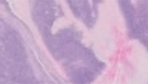
Histologic sections show a biphasic tumor composed of spindled epithelial cells and occasional polygonal epithelial cells admixed with equal proportion of lymphocytes. These cells are arranged in large nodules that are separated by fibrous bands. Mitosis and apoptosis are frequent, imparting a starry-sky appearance. Immunostains are performed and they show the following: The epithelioid cells are positive for cytokeratin. A small area with B-cell follicles is noted. Most lymphocytes are T-cells (CD3 positive) and variably express nuclear TdT. Variable expression of CD4 and CD8 (smear pattern) is also noted, with some cells expressing both CD4 and CD8. In several areas, the tumor cells are present within the capsule and invade through the capsule.
Sections from the adjoining lymph node show anthracosis, sinus histiocytosis, and few non-caseating granulomas that are negative with AFB & GMS stains.
This is a tumor of thymic origin. The flow cytometry profile of CD10 positive T-cell population with variable CD4 and CD8 is characteristic of thymic lymphocytes, in the process of early maturation. Invasion through the capsule with extra-capsular foci makes this a thymoma with some malignant potential. (Of note, the presence of tumor cells outside the capsule may not be represented in all slide sections). The admixture of spindled and rounded epithelial components makes this a type AB thymoma. A mixed lymphocytic and epithelial cell component is also noted.
Thymic neoplasms account for less than 1% of all malignancies with an incidence of approximately 1-5 per million. However, their etiology, biology, and classification are complex. Thymoma is associated with a host of paraneoplastic syndromes, including myasthenia gravis, Good syndrome (combined B- and T-cell immunodeficiency), erythroid hypoplasia, hypogammaglobulinemia, and pure white cell aplasia.
The diagnosis of thymomas entails histological classification (cytological components), assessing the malignant potential (cytologic grade and invasiveness), and TNM staging. Thymomas originate from thymic cortical and medullary lympho-epithelial cells. While the classification of thymomas has evolved frequently and over the years, the current World Health Organization (WHO) system of classification takes into account the cytologic components and their features. Thymomas A, B, and AB exhibit organo-typic architecture (i.e., they tend to recapitulate the thymus). Those composed predominantly of spindled or oval epithelial cells are termed type A thymoma. Those that have a round epithelioid component are called type B thymoma. Admixture of both cell types constitutes AB type. Furthermore, the presence of epithelial atypia and increasing proportion of lymphocytes will constitute B1, B2 and B3 thymomas; B1 thymomas are predominately lymphocytic, whereas B2 and B3 show mixed epithelial and lymphocytic components. Type B3 thymomas tend to have epithelial atypia and display more aggressive biologic behavior. The previous category of thymoma type C does not exist in the current WHO, and these tumors are now reclassified as thymic carcinoma.
Lymphocytes (T-cells) mature in the thymus. They express TdT (an enzyme responsible for nucleotide transfer) within the thymus. The differentiation of a naïve T-cell to a helper (CD4) or cytotoxic (CD8) phenotype occurs within the thymus. During this maturation process, they also express CD10. Once differentiated, almost all normal mature T-cells are either CD4 or CD8 positive, but never express both. Clonal T-cell proliferations (T-cell leukemia and lymphoma) including T-lymphoblastic leukemia/lymphoma (T-ALL), often present with a mediastinal mass, representing a clonal T-cell proliferation within the thymus. Morphologically, the tissue sections of T-ALL reveal only lymphoblasts. The admixed thymic epithelial cell demonstrates minimal if any proliferation. By flow cytometry, the T-ALL may have dual expression of CD4/CD8, TdT and CD10 – similar to the lymphoid population within a thymoma. However, unlike thymoma, these lymphoblasts have monotypic (no variability) expression patterns of the various antigens. This feature can be very useful in some confusing cases of thymoma showing a predominance of lymphoid components.
Malignant mediastinal germ cell tumors comprise up to 5% of mediastinal masses. They are most commonly teratomas (which are histologically similar to teratomas in the gonads) and seminomas which are comprised of larger cells in sheets that are polygonal, have a clear cytoplasm and vesicular chromatin. Lymphoid cells are often seen within the stroma. An organotypic pattern as seen in a majority of thymomas is not seen in germ cell tumors.
Primary complex (primary pulmonary tuberculosis; Ghon complex) patients present with symptoms of lung disease (cough, shortness of breath). The Ghon complex consists of a primary pulmonary tuberculous calcified nodule (usually upper lobe) along with mediastinal hilar lymph node with mycobacterial tuberculosis in a young patient without prior immune exposure to mycobacterium. The appearance of a peripheral lung nodule, prominent lymphatic channels and hilar lymph nodes on radiography was classically shown to be the Ghon complex. The lymph nodes of Ghon focus will show evidence of tuberculosis, which includes granulomatous infection with caseous necrosis and acid-fast bacilli. This case did have associated lymph nodes from the mediastinum (not seen in the slide), but there was no evidence of tuberculous infection. In addition, there was no evidence of pulmonary involvement.
Thymic B-cell hyperplasia is also an uncommon reason for mediastinal masses. This is distinguished by the presence of B-cell hyperplasia (follicles) and relatively preserved T-cells and thymic epithelium. This lesion is seen in over half of all patients with myasthenia gravis.
The classification of thymoma has evolved over several decades and represents the complex biology and variability in morphology this organ and that of tumors originating in this organ. European, Japanese and American pathologists have been varied in their approach and perception of how best to classify thymic tumors. The WHO classification in 1999 which is adopted by the present WHO reflects some of the European based classification systems. The premise of the European pathologists is to reflect biological features and provide histogenetic origin of the tumors (thymic cortex versus medullary origin). While this may hold clinical value, the validation of this system and its use in clinical practice is somewhat limiting. In addition, there is no specific set of immunohistochemical markers or genetic markers readily available to sub-type thymomas based on histogenesis. Thus, even though the current WHO system is effective especially in identifying tumors with aggressive behavior, the classification system can be expected to be revised in the future.
Supplementary Questions
- Which of the following criteria can be used to differentiate T-lymphoblasts in T-lymphoblastic leukemia/lymphoma from T-cells in a thymoma?
- Expression of MPO by T-lymphoblastic leukemia/lymphoma lymphoblasts
- Lack of CD10 expression in T-lymphoblastic leukemia/lymphoma
- T-lymphoblastic leukemia/lymphoma is always negative for CD4 and CD8.
- TdT is expressed only by thymoma T-cells.
- Thymoma T-lymphocytes demonstrate variable expression (smear pattern) of CD4 and CD8 antigens.
- In which of the following features does type A thymoma differ from type B1 and B2 thymoma?
- The presence of atypia
- Thymic epithelium in type A thymoma is rounded.
- Thymic epithelium in type A thymoma is spindled.
- Type A thymoma shows lymphocytic atypia.
- Type B1 and B2 thymomas contain few lymphocytes.
- In which of the following ways do type B3 thymomas differ from type A, B1, and B2 thymomas?
- Type B3 thymomas are the same as thymic carcinoma.
- Type B3 thymomas contain elements of Type A thymomas and Type B thymomas.
- Type B3 thymomas display a spindle cell morphology and less aggressive behavior.
- Type B3 thymomas display epithelial atypia.
References
- Kim, DJ. Prognostic and clinical relevance of the World Health Organization schema for the classification of thymic epithelial tumors: a clinicopathologic study of 108 patients and literature review. Chest. 2005;127(3):755-761.
- Li S, Juco J, Mann KP, Holden JT. Flow cytometry in the differential diagnosis of lymphocyte-rich thymoma from precursor T-cell acute lymphoblastic leukemia/lymphoblastic lymphoma. Am J Clin Pathol. 2004.121(2):268-74.
- Masaoka A, Monden Y, Nakahara K, Tanioka T. Follow-up study of thymomas with special reference to their clinical stages. Cancer. 1981;48(11):2485-2492.
- Moran CA. Weissferdt A, Kalhor N, et al. Thymomas I: A clinicopathologic correlation of 250 cases with emphasis on the World Health Organization schema. Am J Clin Pathol. 2012;137(3):444-450.
- Suster S, Moran CA. "Histologic classification of thymoma: the World Health Organization and beyond." Hematology/Oncology Clinics of North America. 2008; 22(3):381-392.
- Swerdlow SH, Campo E, Harris N, et al, eds. World Health Organization Classification of Tumours of Haematopoietic and Lymphoid Tissues. Lyon, FR: IARC Press; 2008.
- Travis WD, Brambilla E, Muller-Hermelink HK, Harris CC, eds. WHO classification of tumours; Pathology and Genetics of Tumours of the Lung, Pleura, Thymus and Heart. Lyon, FR: IARC Press; 2004.
Author
2016
David Suster, MD
Pathology Resident
Beth Israel Deaconess Medical Center,
Boston, MA
Rajan Dewar, MD, PhD
Surgical Pathology Committee
Beth Israel Deaconess Medical Center and Harvard Medical School
Boston, MA
Answer Key
- Thymoma T-lymphocytes demonstrate variable expression (smear pattern) of CD4 and CD8 antigens. (e)
- Thymic epithelium in type A thymoma is spindled. (c)
- Type B3 thymomas display epithelial atypia. (d)