Clinical Summary
A 24-year-old woman presents with abdominal fullness and a palpable mass on bimanual exam. Imaging reveals a solid mass in the left ovary. Non-enhancing magnetic resonance imaging (MRI) shows high signal intensity centrally with a decreased peripheral signal. At laporatomy, a grey-white bosselated mass with a smooth surface is removed by enucleation. The tumor is yellow-white on sectioning and is largely solid with a few small cystic areas. It measures 12 x 10 x 7 cm in maximum dimensions.
Master List
- Brenner tumor
- Fibroma
- Granulosa cell tumor, adult type
- Krukenberg tumor
- Sclerosing stromal tumor
Archive Case and Diagnosis
This case first appeared as Performance Improvement Program in Surgical Pathology (PIP) 2015, case 10, and is a hepatic nuclear factor 1 alpha (HNF1A)-inactivated hepatocellular adenoma.
Criteria for Diagnosis and Comments
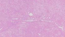
Many of the slides contain hepatic parenchyma composed of banal appearing hepatocytes in a background of macrovesicular steatosis. The sections with adjacent uninvolved liver demonstrate the lesion to have a lobulated contour. Within the mass there are isolated thin-walled arteries and no portal tracts or fibrous septae. There is no mitotic activity, significant inflammation, vascular invasion, pseudoglandular growth pattern or small cell change. Some sections demonstrate focal hemorrhage. There is no significant sinusoidal dilatation. These histologic findings in conjunction with the loss of liver fatty acid-binding protein (L-FABP) by immunohistochemistry (IHC) are diagnostic of hepatic nuclear factor 1 alpha (HNF1A)-inactivated hepatocellular adenoma (HCA).
In its simplicity HCA are benign neoplasms that arise in a background of unremarkable liver composed of normal appearing hepatocytes. Until recently HCAs were thought to be a homogenous entity. However, molecular analysis has changed our understanding of HCAs leading to further subclassification along with raising new questions. Currently in the World Health Organization 2010 classification of HCAs are classified into four categories based on the work of Bioulac-Sage et al. This classification is based on molecular and/or immunohistochemical differences that correlate with the different histologies. The four subtypes are HNF1A-nuclear inactivated HCAs, inflammatory HCAs, beta-catenin-activated HCAs and a small group of HCAs with no defining features. However histologically different these entities are, all contain isolated arteries without associated bile ducts and no thickening of cell plates (best elucidated by evaluating a reticulin stain).
Our case is best classified as an HNF1A-inactivated HCA. This entity is defined by HNF1A biallelic inactivation. These mutations are predominantly somatic. Histologically nearly all of them have macrovesicular steatosis, rarely have inflammation and cytologic abnormalities are uncommon. When evaluating the interface with normal liver they have a lobulated contour. By IHC these HCAs show loss of L-FABP labeling. This can be a critical finding because L-FABP is positive in normal liver and other entities that come up in the differential diagnosis. L-FABP labeling is usually diffuse but may be faint.
HNF1A-inactivated HCAs most commonly develop in women of reproductive age (between ages 20-39) using steroid based oral contraceptives. The incidence of HNF1A-inactivated HCA is greatest in women with long term (greater than 5 years) steroid based oral contraceptive use. It is extremely rare for HNF1A-inactivated HCAs to develop in women who don’t use steroid based oral contraceptives. With the increased use of low dose estrogen oral contraceptives the overall incidence has been declining. The mechanism of action of steroid based oral contraceptives in a development of HCA is not definitively known, however experimental evidence suggests that contraceptives play a role as promoters not initiators. HNF1A-inactivated HCAs are rarely associated with hepatocellular carcinoma (HCC).
Beta-catenin HCAs occur in approximately 40% of men. Beta-catenin HCAs are associated with anabolic/androgenic steroid use as well as non-hormonal risk factors such as glycogen storage diseases. These lesions have activating beta-catenin mutations. This mutation results in nuclear positivity for beta catenin by IHC. One third are associated with cytologic abnormalities (small cell change and pseudoglands) and one fourth have macrovesicular steatosis. Beta-catenin HCAs are diffusely positive for glutamine synthetase whereas normal liver only perivenular areas are positive. These HCA are associated with HCC transformation. In fact recent studies done by Kakar et al suggest that these lesions when occurring in males over 50 may represent low grade HCCs with similar histologic and chromosomal abnormalities.
Inflammatory HCAs are characterized by inflammation (focal or diffuse), sinusoidal dilation, thick walled arteries often associated with a ductular reaction. These lesions are diffusely positive for serum amyloid A (SAA) by IHC. They are most commonly found in women and are associated with diabetes and obesity. They are associated with increased C-reactive protein levels. A small subset of these tumors contains beta-catenin activating mutations. The risk of HCC is rare but those with beta-catenin activating mutations seem to have a higher risk. Unclassified HCAs have no defining histologic or molecular characteristics.
Pathologists making diagnosis of HCA may not have routine access to some of the IHC described above. However understanding of the clinical scenario in conjunction with a careful histologic examination and a “trusty” reticulin special stain should allow one to at least speculate on the specific subtype of HCA. This is of importance because the specific HCA subtype may have a different prognosis and/or treatment.
Clinically most HCAs come to attention incidentally however some present with abdominal pain, an abdominal mass, abnormal liver tests and intraperitoneal hemorrhage. Treatment depends on the HCA subclassification and clinical findings. Clinically significant hemorrhage and the risk of HCC are greatest in tumors over 5 cm. Most commonly HCA are resected, however annual surveillance can be considered in small HCAs (less than 5 cm). Cessation of steroid based oral contraceptives (if taken) usually results in the decrease in HNF1A-inactivated HCAs size. Surgical resection should be undertaken in all beta-catenin activated HCAs. Liver transplantation can be performed in individuals with adenomatosis (greater than 10 adenomas) or unresectable tumors.
Other non-HCAs are in the differential diagnosis in this case. Focal nodular hyperplasia (FNH) are non encapsulated lesions that contain a central or eccentric stellate scar with radiating finger like projections containing large dystrophic vessels and a bile ductular reaction often with accompanying inflammation. These lesions are comprised of unremarkable hepatocytes with no cell plates beyond two cells wide. Importantly these lesions demonstrate “map-like” positivity with glutamine synthetase by IHC meaning there are large anastomosing areas of positivity often around central veins. Focal nodular hyperplasia when well developed has a classic radiologic finding due to the central scar that may negate the need for biopsy. In addition FNHs has early homogeneous enhancement on CT scan which can be diagnostic.
If quickly reviewing a core biopsy of steatotic liver one may go down the wrong “tract”. One must look for portal tracts or isolated vessels. If not realized the pathologist may wrongly assume the target lesion was not sampled and make the diagnosis of steatosis or steatohepatitis. Steatohepatitic HCC is also an entity to be considered. These lesions by definition have macrovesicular steatosis, balloon cells +/- Mallory-Denk bodies, inflammation and pericellular fibrosis. Well to moderately differentiated steatohepatitic HCC may be difficult to differentiate from a HNF1A-inactivated hepatocellular adenoma and this is where a reticulin stain helps and/or IHC for L-FABP. Fibrolamellar HCC may also come up in the differential diagnosis because of the clinical presentation but histologically HCAs do not have the large eosinophilic polygonal hepatocytes with vesicular nuclei and prominent nucleoli, and lamellar fibrosis that characterize fibrolamellar HCC.
Supplementary Questions:
- Which of the following is not associated with an increased risk of hepatocellular adenoma?
- Steroid based oral contraceptive use
- Anabolic/androgenic use
- Glyocogen storage disease
- Obesity
- Microsatellite instability
- Which have the following is not a subtype of hepatocellular adenoma?
- Inflammatory hepatocellular adenoma
- Beta-catenin inactivated hepatocellular adenoma
- Hepatic nuclear factor 1 alpha (HNF1A)-activated hepatocellular adenoma
- Unclassified hepatocellular adenoma
- BRAF mutated hepatocellular adenoma
- Histologically hepatic nuclear factor 1 alpha (HNF1A)-inactivated hepatocellular adenoma typically have which one of the following features?
- Central scar
- Ductular reaction
- Sinusoidal dilatation
- Diffuse steatosis
- Diffuse expression nuclear expression of glutamine synthetase by immunohistochemistry
References
- Bioulac-Sage P,Rebouissou S, Thomas C, et al. Hepatocellular adenomasubtype classification using molecular markers and immunohistochemistry. Hepatology. 2007;46(3):740-748.
- Bosman, F, Carneiro F, Hruban R, Theise N. World Health Organization Classification of Tumours of Digestive System. Lyon, France: IARC;2010.
- Burt A, Portmann B, Ferrell L. MacSween's Pathology of the Liver, 6th Edition: Expert Consult. London, England: Churchhill Livingstone Elsevier;2012.
- Kakar S, Chen X, Ho C, et al. Chromosomal abnormalities determined bycomparative genomic hybridization are helpful in the diagnosis of atypical hepatocellular neoplasms. Histopathology. 2009;55(2):197-205.
- Shafizadeh N, Genrich G, Ferrell L, Kakar S. Hepatocellular adenomas in a large community population, 2000 to 2010: reclassification per current World Health Organization Classification and results of long-term follow-up. Hum Pathol. 2014;45(5):976-983.
- Shafizadeh N, Kakar S. Diagnosis of well-differentiated hepatocellular lesions: role of immunohistochemistry and other ancillary techniques. Adv Anat Pathol. 2011;18(6):438-445.
- Zucman-Rossi J, Jeannot E, Nhieu JT, et al. Genotype-phenotype correlation in hepatocellular adenoma: new classification and relationship with HCC. Hepatology. 2006;43(3):515-524.
Author
William V. Chopp, MD FCAP
Surgical Pathology Committee
Michigan Pathology Specialists
Grand Rapids, MI
Answer Key
- Microsatellite instability (e)
- BRAF mutated hepatocellular adenoma (e)
- Diffuse steatosis (d)