Clinical Summary
A 19-year-old man with a one-year history of polycythemia vera (with associated JAK2 V617F mutation) presents to the emergency room after experiencing epigastric pain for several days. The patient reports recent weight loss and dark urine but denies bleeding, bruising or fever. Physical examination is remarkable for mild jaundice, abdominal tenderness, and hepatomegaly without swelling of the lower extremities. Blood work reveals elevated bilirubin of 5.4 mg/dL and transaminases with an AST of 1600 unit/L and ALT of 2100 unit/L. MRI displays moderate-to-large volume ascites, marked splenomegaly and an enlarged liver with hypertrophy of the caudate and left lobes and atrophy of the right lobe. Both MRI and Doppler ultrasound detect occlusion of the hepatic veins. Due to progression of symptoms and persistence of elevated liver enzymes, the patient receives a liver transplant.
Master List
- Acute viral hepatitis
- Budd-Chiari syndrome (primary)
- Budd-Chiari syndrome (secondary)
- Chronic viral hepatitis with cirrhosis
- Sinusoidal obstruction syndrome (veno-occlusive disease)
Archive Case and Diagnosis
This case first appeared as Performance Improvement Program in Surgical Pathology (PIP) 2015, case 06, and is Budd-Chiari syndrome (primary).
Criteria for Diagnosis and Comments
Budd-Chiari syndrome refers to the broad clinical spectrum associated with hepatic vein outflow obstruction. The obstruction can occur at any level from small hepatic veins to the junction of the inferior vena cava with the right atrium. Patients classically present with the triad of abdominal pain, ascites, and hepatomegaly. Splenomegaly is also common. Budd-Chiari syndrome can be categorized as either primary (intrinsic intraluminal obstruction/thrombosis) or secondary (extravascular origin with intraluminal invasion or obstruction). Primary disease is typically caused by inherited (eg, factor V Leiden, prothrombin gene mutations) or acquired (e.g. myeloproliferative neoplasms, including polycythemia vera, paroxysmal nocturnal hemoglobinuria, oral contraceptive use) hypercoagulable states with resultant thrombosis. Secondary disease can be caused by luminal invasion or compression by neoplasms, abscesses, or cysts.
The underlying etiology of hepatic venous outflow obstruction is usually unknown at the time of initial clinical presentation and requires imaging studies. Presentation can be classified as asymptomatic, fulminant, acute, subacute, or chronic. Obstruction of a single main hepatic vein is clinically silent, whereas the fulminant form typically ensues after rapid and complete obstruction of all main hepatic veins (as in this case). Interestingly, 58% of patients with an acute clinical onset have liver fibrosis, suggesting longstanding disease.
Liver biopsies from patients with suspected or confirmed Budd-Chiari syndrome are uncommon specimens for pathologists given the sensitivity and specificity of radiologic studies. However, liver biopsy is recommended for patients under consideration for a shunting procedure or liver transplant. Venular thrombosis is rarely appreciated histologically with such small sample sizes, but indirect evidence of venous outflow obstruction is typically observed, supported by sinusoidal congestion, hepatocyte necrosis, and fibrosis in the centrilobular areas.
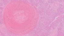
The histologic findings in explanted livers can vary based on duration of disease. Acute lesions, as noted in this case, include dilatation of veins and sinusoids with variable degrees of congestion and necrosis. Acute or organizing thrombi can be seen within the hepatic vein branches, and sometimes within portal veins (most sections in this case showed thrombi within the hepatic vein branches and some slides also showed thrombi in portal veins). As the disease progresses, the sinusoids become collagenized and there is loss of pericentral hepatic parenchyma with replacement by fibrosis. This pattern of fibrosis has been referred to as “congestive cirrhosis” or “venocentric cirrhosis”, in which fibrous bands connect central veins; periportal hepatocytes survive because of retrograde portal vein drainage. In chronic cases of Budd-Chiari, however, it is not uncommon for the more typical “venoportal” pattern of fibrosis (most commonly seen in chronic viral, autoimmune, alcoholic, and biliary disease) to emerge, in which portal tracts are incorporated into the fibrous septae. This would indicate a chronic combined hepatic and portal vein injury. Histologic examination of the hypertrophied caudate lobe may reveal little pathology, as this lobe is often spared from the outflow obstruction due to direct drainage into the inferior vena cava. Benign liver cell nodules (regenerative nodules, focal nodular hyperplasia-like nodules, or adenoma-like nodules) have been observed in non-cirrhotic liver conditions related to vascular abnormalities and are often present in Budd-Chiari syndrome.
Other conditions in the differential diagnosis for suspected hepatic venous outflow obstruction include sinusoidal obstruction syndrome (veno-occlusive disease) and cardiac congestive hepatopathy. These conditions, as well as Budd-Chiari syndrome, can cause similar symptoms and can all result in the histologic findings of sinusoidal congestion and hepatocyte necrosis within centrilobular regions. Correlation with the clinical history and radiologic findings is needed. Sinusoidal obstruction syndrome typically develops within 3 weeks of an acute insult to the sinusoidal endothelial cells. Hematopoietic stem cell transplant, chemotherapy, abdominal radiotherapy, and pyrrolizidine alkaloids are classically associated with the condition. It is thought that the sinusoidal endothelial cells are particularly susceptible to toxic substances because they inherently contain lower glutathione levels compared to neighboring hepatocytes. As a result, the metabolically active zone 3 of the hepatic lobule is most affected. Small hepatic (central) veins containing macrophages and loose fibrosis or concentric intimal/obliterative fibrosis may be noted (and highlighted with a reticulin stain); zone 3 atrophy and sinusoidal fibrosis may also be noted. Congestive hepatopathy results from outflow obstruction related to congestive heart failure or constrictive pericarditis and results in the characteristic gross appearance of a nutmeg liver. Red areas are caused by sinusoidal congestion/bleeding in necrotic regions of lobules, whereas yellow/tan regions correspond to regions of fatty or normal liver tissue. Histologically, congestive hepatopathy may look identical (at least focally) to Budd-Chiari syndrome with dilatation of sinusoids, perivenular hepatocyte necrosis and fibrosis. The presence of hepatic vein thrombi, even in small biopsies, favors Budd-Chiari syndrome. In congestive hepatopathy, hepatic vein edema or fibrosis can be noted and should not be confused with an old or healed thrombus.
Patients with acute viral hepatitis can present with fulminant hepatitis including symptoms of abdominal pain, weight loss, dark urine and jaundice with elevated bilirubin and transaminases. However, portal hypertension and ascites do not typically develop in acute hepatitis and viral serologies should be positive. Histologically, there is severe bridging necrosis with an absence of vascular outflow abnormalities. Conversely, patients with chronic viral hepatitis and cirrhosis can present with elevated liver enzymes, ascites and evidence of portal hypertension, but radiographically and histologically, there would be evidence of cirrhosis with positive viral serologies. Histologically, chronic inflammation within the portal tracts with interface activity would typically be present.
Supplementary Questions:
- A patient presents with acute onset of fatigue, jaundice, and abdominal pain. On imaging, the caudate lobe is hypertrophic and the inferior vena cava appears compressed. The patient’s transaminases continue to rise. A liver biopsy is performed and demonstrates dilated sinusoids with areas of centrilobular hemorrhage and necrosis; the portal tracts show mild chronic inflammation without increased fibrosis. Which of the following is the least likely etiology for this patient’s liver disease?
- Budd-Chiari syndrome (primary)
- Budd-Chiari syndrome (secondary)
- Chronic viral hepatitis with cirrhosis
- Congestive hepatopathy
- Sinusoidal obstruction syndrome (veno-occlusive disease)
- Which of the following statements regarding Budd-Chiari syndrome is true?
- Central veins may contain macrophages or fibrosis that obliterate the lumens.
- Isolated portal vein thrombosis is a well-described feature.
- Patients with chronic venous outflow obstruction involving the hepatic veins often develop bridging necrosis.
- Venocentric hepatocyte dropout and fibrosis is common.
- Zone 1 hepatocytes are at greater risk for necrosis than Zone 2 or Zone 3 hepatocytes.
- Which statement is incorrect?
- Hypercoagulable states are a common cause of Budd-Chiari syndrome.
- Imaging studies (especially Doppler ultrasonography) are an essential part of the workup of Budd-Chiari syndrome.
- Patients with chronic Budd-Chiari syndrome are at increased risk of developing hepatocellular carcinoma.
- Patients with known cardiac disease could present with symptoms of Budd-Chiari syndrome.
- Vascular injury and stasis can cause Budd-Chiari syndrome.
References
- Bayraktar UD, Seren S, Bayraktar Y. Hepatic venous outflow obstruction: Three similar syndromes. World J Gastroenterol. 2007;13(13):1912-1927.
- Burt A, Portmann B, Ferrell L. MacSween's Pathology of the Liver. 6th Ed. Philadelphia, PA: Churchill Livingstone; 2012;601-643.
- Cazals-Hatem D, Vilgrain V, Genin P, Denninger MH, Durand F, Belghiti J, Valla D, Degott C. Arterial and portal circulation and parenchymal changes in Budd-Chiari syndrome: a study in 17 explanted livers. Hepatology. 2003;37(3):510-519.
- Ibarrola C, Castellano VM, Colina F. Focal hyperplastic hepatocellular nodules in hepatic venous outflow obstruction: a clinicopathological study of four patients and 24 nodules. Histopathology. 2004;44(2):172-179.
- Plessier A, Valla DC. Budd-Chiari Syndrome. Semin Liver Dis. 2008;28(3):259-269.
- Tanaka M, Wanless IR. Pathology of the liver in Budd-Chiari syndrome: portal vein thrombosis and the histogenesis of veno-centric cirrhosis, veno-portal cirrhosis, and large regenerative nodules. Hepatology. 1998;27(2):488-496.
Authors
David Martin, MD
Pathology Resident, PGY-3
Emory University Hospital
Atlanta, GA
Alyssa M. Krasinskas, MD
Surgical Pathology Committee
Emory University Hospital
Atlanta, GA
Answer Key
- Chronic viral hepatitis with cirrhosis (c).
- Venocentric hepatocyte dropout and fibrosis is common (d).
- Patients with chronic Budd-Chiari syndrome are at increased risk of developing hepatocellular carcinoma (c).