A 37-year-old woman presents with recent onset of flank pain and a 2-day history of gross hematuria. Urinalysis shows 4+ red cells and red cell casts but no epithelial cells. On radiologic imaging, a heterogeneous mass is noted to displace the left renal pelvis with evident involvement of the parenchyma. Attempted fine-needle aspiration (FNA) yields organized thrombus and fat necrosis with trace mesenchymal cells. Left nephrectomy reveals a 10.0 cm unencapsulated grey and yellow lobulated mass with areas of organizing hemorrhage. The mass arises adjacent to the hilum with displacement of the central calyces, and there is apparent renal parenchymal invasion with islands of parenchyma entrapped by tumor. Microcyst formation is evident.
Master List of Diagnoses
- Angiomyolipoma
- Hemangioma
- Leiomyoma
- Leiomyosarcoma
- Sarcomatoid renal cell carcinoma
Archive Case and Diagnosis
This case first appeared as Performance Improvement Program in Surgical Pathology (PIP) 2016, case 04, and is an angiomyolipoma.
Criteria for Diagnosis and Comments
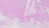
Sections of tumor show mature smooth muscle, aggregates of vascular elements, and mature adipose tissue, which vary in proportion from slide to slide with the muscle fibers in some foci appearing to whorl outward from the vessel walls. The findings are typical of angiomyolipoma (AML). The clinical presentation is relatively classic, as these tumors are typically clinically silent until there is hemorrhage into the renal parenchyma or a calyx; this can be so severe as to be fatal in rare cases, but typically the patient develops flank pain and gross hematuria. Grossly, the tumors may extend into and entrap islands of normal renal parenchyma, with concomitant tubular cysts. There is no evident racial predilection for AML but patients are predominately female with a male-female ratio of 1:2. AML has been described in all age groups but the average age of presentation centers on the 4th and 5th decades.
A classic AML is fairly easy to diagnose on routine hematoxylin and eosin-stained sections, particularly when evaluated in conjunction with clinical history; ancillary stains are not needed in most cases. Many patients have other stigmata of tuberous sclerosis complex (TSC, discussed below). The smooth muscle component is typically bland and derives from the vascular elements of the tumor, with many foci giving a ‘hair on end’ appearance to the muscle fibers as they radiate from the vessels. The vessels are dystrophic muscular arteries with variably-thickened walls, which may blend into more fibrous-appearing areas. Normal elastic lamina is typically absent, or, if present, appears fractured or incomplete; this can be appreciated best with elastin stains. The irregular shape of vessels can lead to small aneurysms which may rupture and precipitate the hemorrhagic presentation of these tumors in some cases.
AMLs were originally considered a benign hamartoma of kidney but immunohistochemical and molecular studies over the past 25 years have resulted in a complete reclassification of this neoplasm, now included in the category perivascular epithelioid cell tumors (PEComa), an expanding group that includes clear cell sugar tumors and lymphangioleiomyomatosis (LAM) of the lung. PEComas have been described in other anatomic locations (gastrointestinal, retroperitoneal, and uterine), typically are spindle cell neoplasms, and derive from a cell with no known normal tissue counterpart, the eponymous ‘perivascular epithelioid cell, (PEC). Eighty percent of patients with TSC manifest AML (often multiple and bilateral), but only 40-50% of patients presenting de novo with AML have additional stigmata of TSC. TSC includes the following physical manifestations: cerebral cortical dysplasia, subependymal giant cell astrocytomas, cutaneous hamartomas, ‘shagreen patches’ (areas of thick, leathery skin typically on the neck), cutaneous hypopigmentation, subungual fibromas or other cutaneous angiofibromas, LAM, and cardiac rhabdomyomas.
Tuberous sclerosis is an autosomal dominant genetic syndrome resulting from mutations of TSC genes 1 and/or 2. TSC1 (9q34) encodes hamartin and TSC2 (16p13) encodes tuberin. TSC1 and TSC2 function as tumor suppressor genes, and while the interactions of hamartin and tuberin remain poorly understood, they are integral to cell growth, division, and cellular interaction. The involvement of two distinct genetic loci is felt to explain the widely variable penetrance of TSC. Mutations of TSC2 appear integral to the expression of LAM but the precise mechanism of action is currently unknown.
The immunohistochemical profile of AML is identical to other PEComas, with diffuse expression of melanocytic markers HMB-45, Melan-A (MART-1), micropthalmia transcription factor (MiTF), and tyrosinase, as well as CD117 (KIT), and without expression of keratins. The constituent elements of AML will also express typical antigens associated with smooth muscle and vascular tissue, such as smooth muscle actin, desmin, and CD34.
The prognosis of AML is ultimately related to the expression of other TSC-associated stigmata, if present. Excision of AML is curative as the classic tumors are not malignant, although rare examples of sarcomatoid variants with more aggressive behavior have been reported. Patients with TSC may have multiple and recurrent AML, in addition to recurrence of other stigmata of the complex. Epithelioid AML is a prominent, but rare, malignant variant. In these tumors, the smooth muscle component is composed of mononuclear epithelioid cells with rounded prominent eosinophilic cytoplasm. Typically, there is some degree of nuclear atypia with pleomorphism and mitotic activity, and the presence of necrosis portends more aggressive behavior, typically local invasion with regional or distant metastasis to liver or lung. The long-term survival for patients with epithelioid AML hovers around 50%.
In its classic form, AML is unlikely to be confused with other renal neoplastic entities. Relative proportions of the 3 major mesenchymal elements may vary from tumor to tumor, and those with a preponderance of one tissue type may prompt consideration of alternative diagnoses.
Renal hemangiomas are rare and may present at any age from youth to late adulthood. As with AMLs, the presenting symptoms are usually gross hematuria and flank pain. Imaging reveals a hyper-echoic mass, and AML is frequently the major differential diagnosis on radiologic study. Phleboliths may be seen. Most hemangiomas arise in or near the renal hilum although they may involve the renal cortex or the papillae. Definitive diagnosis requires excision, and careful histologic exam is required to ensure the absence of the other two tissues types in the AML triad. Not infrequently, patients have other soft tissue or visceral hemangiomas upon more extensive evaluation.
Leiomyomas of the kidney are extremely rare, are derived from the renal capsule, and most typically are incidental autopsy findings, with a size range of 1-5 mm. Symptomatic lesions are quite rare and it is believed that some reported symptomatic larger examples may have been misdiagnosed AMLs with smooth muscle preponderance. Intriguingly, renal capsular leiomyomas reportedly express HMB-45 in addition to conventional smooth muscle markers further suggesting a relationship to AML.
Leiomyosarcoma may be a consideration in cases with increased mitotic activity, atypia, and necrosis. Although some AMLs may manifest focal mitotic activity or ‘atypia’, classic tumors do not express other features of malignancy. As previously mentioned, epithelioid leiomyosarcoma arising in conjunction with the other tissue types, with prominent rounded cells, nuclear atypia, and necrosis is a rare and potentially lethal malignant variant of AML.
Sarcomatoid renal cell carcinomas are carcinomas with a malignant spindle cell component, and this feature portends aggressive behavior and a poor prognosis. There are typically other features of malignancy, with wide regional invasion and necrosis. As most cases have some foci of renal cell carcinoma with more classic features, although it is typically poorly differentiated, confusion with AML is unlikely.
Supplementary Questions:
- Which pair of immunomarkers is most apt to be expressed in both renal angiomyolipomas and lymphangioleiomyomatosis?
- CD10, KIT
- HMB-45, Melan-A
- KIT, CK 5/6
- MART-1, CD10
- Tuberous sclerosis complex has variable penetrance. What is the most likely explanation for the varying degrees of expression of associated stigmata?
- Tuberous sclerosis expression is related to hormone sensitivity to testosterone.
- Tuberous sclerosis is autosomal dominant.
- Tuberous sclerosis is autosomal dominant and controlled by two different genes.
- Tuberous sclerosis is sex-linked.
- Gross hematuria and flank pain are common presenting signs and symptoms of angiomyolipoma and which of the following?
- Hemangioma
- Leiomyoma
- Leiomyosarcoma
- Lipoma
References
- Bissler JJ, Henske EP. Renal Manifestations of Tuberous Sclerosis Complex. In: Kwiatkowski DJ, Whittlemore DJ, Thiele EA, eds. Tuberous Sclerosis Complex: Genes, Clinical Features and Therapeutics. Weinheim, DE. Wiley-VCH Verlag GmbH; 2010:321–325.
- Hornick, JL, Fletcher CD. PEComa: What do we know so far? Histopathology. 2006;48(1):75-82.
- Murphy WM, Grignon DJ, Perlman EJ. Tumors of Kidney, Bladder, and Related Urinary Structures. Atlas of Tumor Pathology. Series 4, Fascicle 1. Washington, DC: American Registry of Pathology/ Armed Forces Institute of Pathology; 2004:187-194.
- Rakowski SK, Winterkorn EB, Paul E, Steele DJ, Halpern EF, Thiele EA. Renal manifestations of tuberous sclerosis complex: Incidence, prognosis, and predictive factors. Kidney Int. 2006;70(10):1777-1782.
Author
2016
Philip A Branton, MD
Surgical Pathology Committee
Biorepositories and Biospecimens Research Branch, National Cancer Institute
Rockville, MD
Answer Key
- HMB-45, Melan-A (b)
- Tuberous sclerosis is autosomal dominant and controlled by two different genes. (c)
- Hemangioma (a)