- Home
- Member Resources
- Pathology Case Challenge
- Inguinal Lymph Node
Clinical Summary
A 28-year-old man presents with generalized adenopathy and weight loss. The patient is otherwise asymptomatic. Peripheral blood examination is normal. A firm mobile inguinal mass that developed over the last six months is removed. Gross examination reveals two fragments of soft tissue (30 and 20 grams), measuring up to 5.0 cm, with a lobulated tan cut surface. Immunophenotypic flow cytometry analysis reveals no clonal proliferation.
Master List
- Blastomycosis
- Granulomatous lymphadenitis/sarcoidosis
- Histiocytic lymphadenopathy with foreign body type giant cell reaction
- Toxoplasmosis
- Tuberculous lymphadenitis
Archive Case and Diagnosis
This case first appeared as Performance Improvement Program in Surgical Pathology (PIP) 2014, case 22 and is a granulomatous lymphadenitis/saccoidosis.
Criteria for Diagnosis and Comments
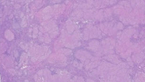
The histological sections reveal fragments of enlarged matted lymph nodes with effaced architecture. Subcapsular sinuses and germinal centers are replaced by tight aggregates of epithelioid histiocytes with occasional giant cells. Some giant cells contain intracytoplasmic grey or dark blue concentric calcifications (calcium oxalate, "conchoidal bodies"). The asteroid bodies are rare. There is no evidence of polarizable foreign matter. Many smaller granulomas are surrounded by a thin rim of lymphocytes and plasma cells. Some granulomas coalesced into larger (subcapsular) nodules with more prominent refractile eosinophilic collagen bands. Very rare tissue sections show small round foci of punctate necrosis. Even when present, the foci of necrosis show no neutrophils. Microabscesses are absent. There is no evidence of lipogranulomatous inflammation. A GMS stain for fungi and AFB stain for acid fast bacilli are negative. If the infectious etiology of this predominantly non-necrotizing granulomatous lymphadenitis is excluded (e.g., negative tissue cultures), the histological presentation is most consistent with that of sarcoidosis. The possibility of sarcoidosis can be further addressed by a combination of clinical, radiographic and laboratory studies (e.g., angiotensin-converting enzyme level).
Sarcoidosis is a multisystem disease and non-necrotizing granulomas may develop in any organ. Most commonly, patients present with pulmonary involvement and intrathoracic lymphadenopathy. However, a subset of patients with sarcoidosis may present with peripheral lymphadenopathy. In the absence of respiratory or cutaneous symptoms, peripheral lymph node biopsy may be supportive of sarcoidosis in up to 66% of cases. Of all peripheral lymph nodes, cervical, supraclavicular, and inguinal appear to be affected most frequently.
The etiology of sarcoidosis is still unresolved. Several epidemiologic observations, including spatial and seasonal clustering of patients, raise the possibility of an infectious agent. Mycobacterial and propionibacterial microorganisms are the most frequently implicated infectious agents. The possibility of latent infections by these organisms complicates the interpretation of most immunologic studies. Currently, Propionibacterium acnes (P. acnes; gram-positive, anaerobic bacterium) is the only microorganism that was isolated by bacterial cultures from up to 78% of lymph nodes with sarcoid granulomas. P. acnes or P. granulosum were detected by polymerase chain reaction, immunohistochemistry, and in situ hybridization in most cases of sarcoidosis and absent from most normal controls and samples of tuberculosis. Importantly, Mycobacterium avium was not detected in cases of sarcoid, while M. tuberculosis was identified in only 0-9% of sarcoid specimens. Interestingly, pulmonary granulomatosis induced by P. acnes antigen in mice and rabbits can be prevented by antibiotics. Antibiotic therapy (minocycline, doxycycline, and clarithromycin) appears to be effective in about 50% of patients with sarcoidosis. However, it is still unclear whether these antibiotics are effective due to their antimicrobial properties or because of their well-recognized anti-inflammatory effect.
In our case, the foci of necrosis are very rare, small, and rounded (rather than irregular). Combined with negative AFB and GMS stains, the possibility of tuberculous or fungal lymphadenitis is unlikely. The lack of microabscesses in small areas of necrosis and negative GMS stain also argue against the possibility of blastomycosis. Nevertheless, correlation with tissue culture results is needed. Toxoplasma gondii usually affects cervical lymph nodes. Histologically, toxoplasma lymphadenitis can be recognized by preserved lymph node architecture with follicular hyperplasia, pale monocytoid cells in nodal sinuses, and epithelioid histiocytes in mantle zones and germinal centers.
In some patients who had undergone hip replacements, microparticles of cobalt-chromium alloy, titanium, or polyethylene can migrate from the prosthesis to ipsilateral inguinal and pelvic lymph nodes and elicit prominent non-granulomatous sinus histiocytosis. Non-necrotizing granulomatous lymphadenitis mimicking sarcoid may also be secondary to an unrecognized malignancy in the region draining into the involved lymph node or due to anti-neoplastic treatment (e.g., interferon, interleukin-2) in a known oncologic patient. There is no history of hip replacement or malignancy in our patient.
Cases of sarcoid-like disease or sarcoidosis have been reported in patients receiving several anti-TNF (tumor necrosis factor) therapeutic agents. This is a rather unexpected complication as some cases with bona fide refractory sarcoid actually respond to anti-TNF therapy. In patients on anti-TNF therapy, dyspnea and/or coughing usually lead to chest radiography, which may reveal newly developed bilateral hilar adenopathy. Although the lungs are most often affected in such scenarios, common extrapulmonary abnormalities include skin lesions. The condition improves with cessation of the anti-TNF agent and/or corticosteroid treatment. There was no history of autoimmune disease or anti-TNF therapy in our patient.
Supplementary Questions:
- Which microorganism is repeatedly isolated by bacterial culture from sarcoid granulomas?
- Epstein-Barr virus
- Escherichia coli
- Mycobacterium avium
- Mycobacterium tuberculosis
- Propionibacterium acnes
- What class of drugs was recently shown to be paradoxically associated with sarcoid-like reaction?
- Antibiotics
- Anti-epidermal growth factor receptor antibodies
- Anti-inflammatory corticosteroids
- Anti-TNF
- Tyrosine kinase inhibitors
- Sarcoidosis is the diagnosis of exclusion. For instance, sarcoid-like reaction to chemotherapy and the possibility of infectious etiology should be excluded before accepting the diagnosis of sarcoidosis.
- True
- False
References
- Albores-Saavedra J, Vuitch F, Delgado R, et al. Sinus histiocytosis of pelvic lymph nodes after hip replacement. A histiocytic proliferation induced by cobalt-chromium and titanium. Am J Surg Pathol. 1994;18(1):83-90.
- Cleynen I, Vermeire S. Paradoxical inflammation induced by anti-TNF agents in patients with IBD. Nat Rev Gastroenterol Hepatol. 2012;9(9):496-503.
- Eishi Y. Etiologic link between sarcoidosis and Propionibacterium acnes. Respir Investig. 2013;51(2):56-68.
- Yanardag H, Caner M, Papila I, et al. Diagnostic value of peripheral lymph node biopsy in sarcoidosis: a report of 67 cases. Can Respir J. 2007;14(4):209-211.
Author
2014
Simion I. Chiosea, MD FCAP
Surgical Pathology Committee
University of Pittsburgh Medical Center
Pittsburgh, PA
Answer Key
- Propionibacterium acnes (e).
- Anti-TNF (d).
- True (a).