Clinical Summary
A 57-year-old Hispanic man had a long history of non-ischemic dilated cardiomyopathy. He grew up on a farm in El Salvador and immigrated to the United States as a young man where he has worked as a manual laborer all of his life. His course was complicated by arrhythmias, progressive intractable ventricular tachycardia and AV node block. Following numerous clinical interventions spanning several years, orthotopic heart transplantation was performed. Four months later, the patient developed severe cardiac failure. A cardiac biopsy was performed and a laboratory assay was obtained, both of which proved to be diagnostic for a disease process. In spite of aggressive therapy, his disease worsened and he died two weeks later. An autopsy was performed. The glass slide provided contains a representative sample of the transplanted heart.
Master List
- Acute cellular rejection, Grade 3R (ISHLT 2004)
- Candida infection
- Chagas disease, acute reactivation following transplantation
- Cytomegalovirus infection
- Toxoplasmosis
Archive Case and Diagnosis
This case first appeared as Performance Improvement Program in Surgical Pathology (PIP) 2013, case 08, and is Chagas disease, acute reactivation following transplantation.
Criteria for Diagnosis and Comments
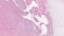
Histologic sections show a dramatic myocarditis spanning the full thickness myocardium and epicardium. Inflammatory cells consist primarily of lymphocytes with fewer numbers of plasma cells, histiocytes and rare eosinophils and neutrophils. Interstitial edema is prominent, and myofiber degeneration is readily identified. Many myocardial fibers contain large numbers of intracellular organisms (amastigote forms) which are aggregated together in dilated cyst-like spaces within the cytoplasm. Tissue Gram, silver and PAS stains do not highlight the organisms. These findings represent infection by trypanosoma cruzi, otherwise known as Chagas disease. The pre-mortem cardiac biopsy similarly showed the organisms, and the diagnostic clinical assay was a serum based ELISA which was positive for Trypanosoma cruzi.
Chagas disease, also called American trypanosomiasis, is a tropical parasitic disease first described in the early 20th century by Brazilian physician Carlos Chagas. Originally studying a malaria outbreak on the Sao Francisco River in 1909, Chagas noted an unusual illness affecting local villagers. He observed infestation of their rural houses with hematophagous (blood sucking) insects which he proved harbor flagellate protozoa in their intestines. Chagas was then able to demonstrate that this newly discovered protozoon could be transmitted to monkeys via infected insects. He named the protozoa T. cruzi, after the Brazilian researcher Oswaldo Cruzi, and demonstrated the armadillo as the natural reservoir. Based on his suspicion that the protozoa were pathogenic, Chagas went on to show that the organism could cause acute parasitemia, myocarditis and meningoencephalitis in humans. He also performed over 100 autopsies on patients with suspected longstanding disease, carefully documenting what would eventually prove to be the pathologic features of chronic Chagas disease.
The mode of entry into humans is typically via direct inoculation of the trypomastigote protozoan form into the bite site, eye or oral cavity. The organism can also be transmitted via blood transfusion, maternal fetal transfer, or rarely via contaminated food products. The acute phase of the disease is usually mild, with swelling at the inoculation site. Parasitemia may be seen in the first few weeks and antiparasitic therapies are successful in eradicating the disease. Rarely, acute myocarditis and meningoencephalitis occur, pathology of which is characterized by active inflammation in reaction to numerous protozoan amastigote forms within tissue. Beyond 4-8 weeks, untreated patients enter the chronic phase of Chagas disease, of which 80% will remain asymptomatic. Over decades, approximately 20% of chronic phase patients will develop clinical symptoms. The pathologic changes are secondary to ongoing inflammation, fibrosis and scarring in reaction to the very low numbers of amastigote forms harbored in tissue. Parasitemia is no longer observed. Organs commonly involved include the brain, colon and esophagus, with resultant encephalitis, megacolon and megaesophagus. Approximately two thirds of these patients will develop cardiac disease which often manifests clinically with arrhythmias. Pathologic findings in the heart include dilated cardiomyopathy, left ventricular apical aneurysm formation, mural thrombi, interstial fibrosis and varying degrees of chronic inflammatory cell infiltrates. Amastigotes are generally difficult (or not possible) to identify microscopically. Diagnosis of the disease in the chronic phase generally rests on serologic immunoassays or PCR, and a limited number of laboratories offer an immunohistochemical tissue assay.
Chagas disease is endemic in Latin American countries, affecting an estimated 10 to 16 million people, where the primary mode of transmission remains the insect vector. Although hematophagous insects carrying T. cruzi are found in the southern United States, the extent of insect transmission appears to be extremely rare. The more important problem of Chagas disease in the USA are the large numbers of immigrants from Central and South America, who have as yet undiagnosed chronic disease, with estimates of around 300,000 people. Increasing numbers of cases of blood and tissue transmission of T. cruzi have occurred, and now most blood and tissue products are screened in the USA. As with the current patient, who proved to be in the latent chronic phase of Chagas disease, immunosuppressive therapies related to transplantation or cancer treatment can cause reactivation as acute phase Chagas disease.
The differential diagnosis of T. cruzi infection in the heart is very limited. In this particular case of heart transplantation, one must consider acute cellular rejection. However, acute cellular rejection generally manifests with influx of lymphocytes and histiocytes, and typically does not have this degree of cellularity. The intracellular organisms are also characteristic of T. cruzi and not cellular rejection. In this clinical setting, one must consider other infectious agents such as Candida, cytomegalovirus and toxoplasmosis. The morphology of Candida and cytomegalovirus are sufficiently unique and are thus generally not easily confused with T. cruzi organisms. Toxoplasma gondii is a parasite that can acutely infect the myocardium causing myocarditis, especially in immunocomprimised patients as in this case. This protozoa has morphology similar to T. cruzi, and will accumulate as intracytoplasmic cysts in myocardial cells, features which make the distinction between the two very challenging. As antibodies exist to both organisms, immunohistochemical assays may be necessary to confirm the diagnosis. Clinical history and serologic assays are also potentially useful in this differential.
Supplementary Questions:
- The organism responsible for causing Chagas disease is
- Cytomegalovirus
- Epstein Barr virus
- Listeria monocytogenes
- Tinea cruris
- Trypanosoma cruzi
- The chronic phrase of T. cruzi infection is characterized by all of the following except?
- Cardiac aneurysm
- Dilated colon
- Few intracellular amastigote forms
- Parasitemia
- Spans many years duration
- Immunosuppression is a risk factor for systemic infection by which of the following:
- Candida
- Cytomegalovirus
- Toxoplasma gondii
- Trypanosoma cruzi
- All of the above
References
- Bern C. Antitrypanosomal therapy for chronic Chagas disease. N Engl J Med. 2011; 364:2527-2534.
- Bern C, Montgomery SP. An estimate of the burden of Chagas disease in the United States. Clinical Infectious Disease. 2009;49(5):e52-54.
- Bern C, Montgomery SP, Herwaldt BI, Rassi A, et al. Evaluation and treatment of Chagas disease in the United States: a systematic review. JAMA. 2007;298(18):2171-2181.
- Benvenuti LA, Roggerio A, Coelho G, Fiorelli AI. Usefulness and qualitative polymerase chain reaction for trypanosoma cruzi DNA in endomyocardial biopsy specimens of chagasic heart transplant patients. J Heart Lung Transplant. 2011;30(7):799-804.
- Chin-Hong PV, Schwartz BS, Bern C, Montgomery SP, et al. Screening and treatment of Chagas disease in organ transplant recipients in the United States: recommendations from the Chagas in transplant working group. Am J Transplant. 2011;11:1-9.
- Leslie M. Drug developers finally take aim at a neglected disease. Science. 2011;333:933-935.
- Voelker R. A century after Chagas disease discovery, hurtles to tackling the infection remain. JAMA. 2009;302(10):1045-1047.
Author
2012
Daniel J. Luthringer, MD
Surgical Pathology Committee
Cedars-Sinai Medical Center
Los Angeles, CA
Answer Key
- Trypanosoma cruzi (e).
- Parasitemia (d).
- All of the above (e).