Clinical Summary
A 68-year-old woman presents with headache and seizures of recent onset. Computed tomography (CT) of the head shows a 4.0 x 3.0 cm contrast-enhancing, well-defined, left parasagittal mass compressing the parietal lobe. The mass is resected and on gross examination shows a firm, well-demarcated tan-white tumor attached to a small segment of dura.
Master List:
- Meningioma
- Meningothelial hyperplasia
- Schwannoma
- Solitary fibrous tumor
Archive Case and Diagnosis
This case first appeared as Performance Improvement Program in Surgical Pathology (PIP) 2015, case 20 and is a meningioma.
Criteria for Diagnosis and Comments
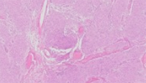
Histologic sections show features typical of the syncytial type of classic meningioma characterized by lobules of uniform cells partly demarcated by thin collagenous septae. The cells have abundant cytoplasm, indistinct cytoplasmic borders, and appear to form a syncytium within the lobule. The nuclei are oval with delicate chromatin and exhibit occasional nuclear cytoplasmic inclusions. Whorl-like arrangement of the cells and psammoma bodies are present in some of the submitted sections.
Meningiomas are generally dura based neoplasms arising from the meningothelial (arachnoid) cells; most are sporadic with a few occurring after irradiation and rarely in association with NF2. Meningiomas account for 24-30% of primary intracranial tumors and occur most commonly in middle aged and elderly patients (peak during the 6th and 7th decades) with a striking female preponderance (3:2). More than 90% of meningiomas occur intracranially typically over the cerebral convexities, often parasagittal in association with the faux and venous sinus. Less commonly, meningiomas arise from the optic nerve, within the spinal cord and the ventricular system. Meningiomas are typically slow-growing, asymptomatic tumors often discovered as incidental findings on neuroimaging. When symptomatic, neurologic signs and symptoms are related to location and compression of adjacent structures and include focal neurologic deficits, increased intracranial pressure, and seizures. Meningiomas arising from the optic nerve cause visual symptoms and those arising within the spinal cord may cause radicular pain. Grade II and III meningiomas cause cerebral edema.
The Word Health Organization (WHO 2007) classification of meningiomas includes 3 grades:
Grade I (benign): most common (80%) and appear as well-circumscribed, firm, white-tan masses, often attached to dura. Infiltration of bone and scalp may occur and calcifications and hyperostosis of bone adjacent to the tumor are frequent findings. Of the many morphologic variants of meningiomas, syncytial (meningothelial), fibrous, and transitional types are the most common. Syncytial type is characterized by monomorphic cells with oval to round nuclei arranged in whorls or lobules within which the cells form syncytia while the fibrous type is characterized by spindled cells arranged in interlacing bundles in a collagenous background. Transitional/mixed type of meningioma shows a pattern that is intermediate between the syncytial and fibrous types or a mixture of syncytial and fibrous patterns. Other variants include psammomatous, angiomatous, microcystic, metaplastic, lymphoplasmacytic, and secretory. Treatment approach and prognosis are essentially similar in all Grade I meningiomas.
.
Grade II (atypical): includes atypical, clear cell, and chordoid meningiomas. Atypical meningiomas have increased mitotic activity (≥4 mitoses per ten high power fields), brain invasion, or at least three of the following five microscopic features: sheet-like growth pattern, nuclei with macronucleoli, hypercellularity, small cell formation, or spontaneous necrosis.
Grade III (anaplastic): includes anaplastic, papillary, and rhabdoid meningiomas. Anaplastic meningiomas have ≥20 mitoses per ten high power fields and/or malignant characteristics resembling carcinoma, sarcoma, or melanoma. Features that support the diagnosis of malignant meningioma include the loss of usual meningioma growth patterns, infiltration of underlying brain, abundant mitoses with atypical forms, and multifocal microscopic foci of necrosis.
By immunohistochemistry, approximately 80% of meningiomas are positive for epithelial membrane antigen (EMA) and claudin, up to 40% are at least focally positive for S-100 protein (fibrous variant more commonly positive), and a minority of tumors are positive for CEA and cytokeratin. A majority of tumors express progesterone receptors but this is less likely in atypical or anaplastic meningiomas. Meningiomas are generally negative for GFAP.
.
MIB-1 labeling index correlates with grade and recurrence rate with an index greater than 4% associated with increased recurrence. Mean values of MIB-1 index: benign, 3.8%; atypical, 7.2%; anaplastic, 14.7%.
By cytogenetics studies, loss of the long arm of chromosome 22, which is usually associated with inactivation of the NF2 gene, is the most common genetic abnormality found in meningiomas. Mutations in the NF2 gene are found in the majority of NF2-associated and up to 60% of sporadic meningiomas. Additional chromosome abnormalities associated with increased grade include loss of heterozygosity for chromosome 1p, loss of 14q, deletion of 9q21, and abnormalities of chromosomes 6q, 10, 14q, and 17q.
The differential diagnoses of meningioma vary by the anatomic location of the tumor and the histologic pattern. Since the lesional cells of meningioma closely resemble the normal arachnoid cap cells, reactive meningothelial hyperplasia may be considered in the histologic differential diagnosis on small biopsy specimens. However, meningothelial hyperplasia is typically associated with a predisposing factor such as hemorrhage, chronic renal failure, or trauma, and is often present adjacent to other neoplasms including gliomas.
Schwannoma is in the histologic differential diagnosis of fibrous meningioma especially when occurring in the cerebellopontine angle. Schwannoma is a biphasic tumor consisting of highly cellular areas (Antoni A) admixed with loose spongy areas of lower cellularity (Antoni B) and typically lacks the distinct whorled architecture and psammoma bodies seen in meningioma. Verocay body formation and diffuse S-100 protein positivity are additional distinguishing features of schwannoma.
Solitary fibrous tumor is characterized by a spindle cell proliferation with numerous variably sized, slitlike and staghorn-like vessels. The lesional cells are negative for EMA, and diffusely positive for CD34.
Distinction of anaplastic meningiomas from other malignancies, including carcinoma, melanoma, or sarcoma, can be challenging and requires correlation of clinical, radiologic, and immunohistochemical findings.
The majority of meningiomas are benign and complete surgical resection typically offers an excellent prognosis with radiation therapy shown to be beneficial in recurrent or unresectable tumors. Benign meningiomas have a recurrence rate of 7% to 25% while atypical meningiomas are associated with a 29% to 52% recurrence rate. Anaplastic meningiomas are associated with poor prognosis related to high recurrence rate (50-95%) and metastatic potential.
Supplementary Questions
- Which of the following immunomarkers is most likely to be positive in meningioma?
- CD117
- Cytokeratin
- EMA
- Estrogen receptors
- GFAP
- Which of the following features is used in characterizing an otherwise benign meningioma (Grade I) as atypical meningioma (Grade II)?
- Brain invasion
- Expression of progesterone receptors
- Psammoma bodies on histology
- Syncytial pattern on histology
- Ventricular location
- Which of the following variants is regarded as atypical (Grade II) meningioma?
- Angiomatous meningioma
- Chordoid meningioma
- Fibrous meningioma
- Papillary meningioma
- Psammomatous meningioma
References
- Alahmadi H, Croul SE. Pathology and genetics of meningiomas. Semin Diagn Pathol. 2011;28(4):314-324.
- Mawrin C, Perry A. Pathological classification and molecular genetics of meningiomas. J Neurooncol. 2012;99:379-391.
- Nakasu S, Li DH, Okabe H, et al. Significance of MIB-1 staining indices in meningiomas. Am J Surg Pathol. 2001;25:472-478.
- Perry A, Louis DN, Scheithauer BW et al. Meningiomas. In: Louis DN, Obgaki H, Wiestler OD, Cavenee WK, ed. WHO Tumours of the Central Nervous System. Lyon, FR: IARC; 2007:164-172.
- Prayson RA. Non-glial tumors. In: Goldblum JR. Neuropathology. Philadelphia, PA: Elsevier; 2012:513-518.
- Vranic A, Peyre M, Kalamarides M. New insights into meningioma: from genetics to trails. Curr Opin Oncol. 2012;24:660-665.
Author
Vijaya B. Reddy, MD
Surgical Pathology Committee
Rush University Medical Center
Chicago, IL
Answer Key
- EMA (c)
- Brain invasion (a)
- Chordoid meningioma (b)