- Home
- Member Resources
- Pathology Case Challenge
- Uterus/Endometrium
Clinical Summary
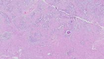
A 56-year-old woman presents with abnormal uterine bleeding. An endometrial biopsy is performed and the diagnosis leads to a decision to perform a hysterectomy.
Master List
- Clear cell carcinoma
- Endometrioid carcinoma
- Mucinous carcinoma/serous carcinoma
Archive Case and Diagnosis
This case first appeared as Performance Improvement Program in Surgical Pathology (PIP) 2014, case 15 and is an endometrioid carcinoma.
Criteria for Diagnosis and Comments
Submitted sections consist of myometrium with an infiltrating neoplasm composed of glandular cells with enlarged round-oval nuclei with clear to granular chromatin and occasional prominent nucleoli. In approximately 30% of the tumor, the cells are growing in solid sheets with loss of gland or acinar formation. Some slides demonstrate foci of squamous metaplasia. There is a desmoplastic stromal reaction, and those slides that include the serosal surface of the uterus show an infiltrating tumor closely approximating the serosa. Lymph-vascular tumor emboli can be seen in subserosal lymphatics in some sections. The histologic features are those of adenocarcinoma of the endometrium, endometrioid carcinoma (EC). The relative proportion of solid areas of carcinoma qualifies for classification as grade 2 by FIGO criteria.
Carcinomas of the endometrium are the commonest malignancy of the gynecologic tract in the USA and Western Europe. They are clinically and pathologically divided into two major groups: type I, which includes endometrioid carcinomas and its variants, as well as mucinous carcinoma; and type II, which includes serous, clear cell, squamous, and undifferentiated carcinomas.
Generally, type I tumors occur in peri-menopausal women, arise from hyperplastic precursor lesions (complex hyerplasia with atypia, for example) in a milieu of unopposed estrogen; and typically, but not universally, are minimally invasive and less aggressive. Type II tumors tend to arise in post-menopausal patients and are not related to unopposed estrogen or a hyperplastic precursor lesion. Aggressive behavior is more common with deep invasion and metastases. On the molecular level, type I tumors show microsatellite instability, KRAS and PTEN mutations, and beta catenin nuclear accumulation. Type II tumors are more apt to show loss of heterozygosity (LOH) at multiple loci and p53 mutations. These are general categories, and exceptions or overlap of features can occur in an individual case, such as the current one, which manifests an endometrioid phenotype but more aggressive behavior with deep myometrial and lymphvascular invasion. For this reason, careful grading and staging of all tumors, regardless of phenotype, is required.
EC is by far the most common variant of endometrial carcinoma, accounting for roughly 80% of all endometrial malignancies. Grading of endometrioid carcinomas assesses the relative percentage of solid or non-glandular, non-squamous, confluent growth to glandular (i.e., better differentiated) portions of the tumor. Tumors with less than 5% solid growth are considered grade 1, 6–50% are grade 2, and more than 50% solid growth qualifies for grade 3. Myometrial invasion by carcinoma is an independent prognostic factor, with those tumors manifesting invasion more than 50% of myometrial thickness having a worse prognosis than those tumors that are confined to the endometrium, or that invade the myometrium less than 50% of thickness.
The presence of lymph-vascular invasion (LVI) is a risk factor for lymph node involvement by metastatic carcinoma, and its presence may prompt a follow-up lymph node dissection or sampling of pelvic and para-aortic nodes, if this was not performed during the original hysterectomy. For this reason, careful evaluation for LVI should be performed on all sections.
A special note should be made of endometrioid carcinomas in the setting of Hereditary Non-polyposis Colon Cancer syndrome (HNPCC), or Lynch syndrome. The relative lifetime risk of endometrial carcinoma is 40–60% in female patients with Lynch syndrome and it actually exceeds the lifetime risk of colon cancer in younger female patients with this syndrome. Lynch syndrome-associated endometrial carcinomas express germline defects in DNA mismatch repair genes, typically MLH1 or MSH2. The tumors classically express some characteristic features including higher tumor grade, marked lymphocytic permeation and/or "Crohn disease-like" lymphoid reaction, and extensive lymph-vascular permeation. The diagnosis of endometrial carcinoma in a pre-menopausal patient, particularly with the above features, should prompt a comment to the clinician to consider evaluation for Lynch syndrome, and immunostaining of the tumor for MLH-1, PMS-2, MSH-2, or MSH-6.
Serous carcinomas (SC) of the endometrium manifest the same cytologic features as those seen in the ovary, with hyperchromatic nuclei and variable amounts of pinkish cytoplasm. Serous carcinoma accounts for 5–10% of cases of endometrial carcinomas and is the prototypic type II endometrial carcinoma. The cells grow at with thick fibrovascular cores with complex arborization, and prominent lining cellular stratification and tufting, with exfoliation or cellular budding in a ‘candle dripping’ pattern. Stromal desmoplasia is typical. The tumors typically are highly aggressive with deep myometrial invasion and LVI; extra uterine metastases or synchronous malignancies of ovary and fallopian tube are common. Serous carcinomas derive from endometrial intraepithelial carcinoma (EIC) in atrophic endometrium or in an endometrial polyp. Cells in EIC are highly atypical, while non-invasive, and cytologically are identical to those cells of invasive SC, are typically strongly reactive with anti-p53 antibodies, and show a high percentage of reactivity with anti-Ki-67 antibodies.
Clear cell carcinoma (CCC) derives it eponymous name from the prominent, optically clear cytoplasm characteristic of the tumor cells, which may manifest intracytoplasmic hyaline bodies as well. It is included in the type II endometrial carcinomas and accounts for approximately 5% of endometrial carcinomas. The nuclei are typically hyperchromatic with prominent nucleoli, and the cells classically form 'hobnail' blunt projections into acinar lumina or along fibrovascular cores, but may also form tubulocystic and solid patterns. Because CCC occasionally appears in conjunction with EC, some authorities have speculated it was a 'forme fruste' of EC, but its lack of ER and PR expression and other studies have shown it is more closely related to serous carcinoma, although it lacks the strong immunoreactivity to p53 antibodies seen in serous carcinoma. As noted previously, CCC is a tumor of the later decades and while the tumor may appear deceptively non-aggressive at presentation, recurrence with metastasis is common.
Mucinous carcinoma (MC) accounts for less than 10% of endometrial carcinomas and is defined by the presence of intracytoplasmic mucin in more than 50% of tumor cells; intra-luminal or acinar mucin is common in endometrioid and other variants and does not qualify a tumor for this designation MC frequently co-exist with conventional endometrioid carcinomas; they commonly resemble endocervical mucinous tumors to some degree and typically are relatively low-grade and low-stage, though some degree of myometrial invasion is not uncommon.
Supplementary Questions:
- Which of the following is true regarding type 1 endometrial carcinomas?
- They are associated with p53 mutations.
- They are typically highly aggressive with deep invasion and widespread metastases.
- They are more common in elderly or postmenopausal patients.
- They are associated with KRAS and PTEN mutations.
- Clear cell carcinoma is a typical example of a type 1 endometrial carcinoma.
- In which patient should a recommendation for evaluation for Lynch syndrome be considered?
- 21-year-old patient with simultaneous squamous carcinoma of cervix and an endometrial polyp
- 32-year-old patient with grade II endometrioid carcinoma with prominent lymphoid tumor response
- 40-year-old patient with noninvasive mucinous carcinoma
- 80-year-old patient with serous endometrial carcinoma
- 90-year-old patient with carcinosarcoma and long history of tamoxifen therapy
- Which of the following is true with respect to the assessment of lymph-vascular invasion (LVI) in endometrial carcinoma?
- Its presence automatically makes the tumor stage IV.
- It has no effect on prognosis.
- It is predictive of lymph node involvement by tumor.
- It is only a factor in type II endometrial carcinomas.
- It is only seen in patients with Lynch syndrome.
References
- Meyer LA, Broaddus RR, Lu KH. Endometrial cancer and Lynch syndrome: clinical and pathologic considerations. Cancer Control. 2009;16(1):14-22.
- Neubauer NL, Lurain, JR. The role of lymphadenectomy in surgical staging of endometrial cancer. Int J. Surg. Onc. 2011. Doi: 10.1155/2011/814649
- Nucci MR, Oliva E. Gynecologic Pathology. New York: Churchill Livingston/Elsevier 2009;240-251.
Author
2014
Philip A Branton, MD FCAP
Surgical Pathology Committee
National Cancer Institute, NIH
Bethesda, MD
Answer Key
- They are associated with KRAS and PTEN mutations. (d).
- 32-year-old patient with grade II endometrioid carcinoma with prominent lymphoid tumor response (b).
- It is predictive of lymph node involvement by tumor. (c).