- Home
- Member Resources
- Pathology Case Challenge
- Thyroid Gland
Clinical Summary
A 48-year-old woman presents with hypothyroidism treated with high dose Synthroid® for the past 28 months. Recently, she has developed marked dysphagia and dyspnea. She has a history of classical Hodgkin lymphoma 20 years ago. Physical exam reveals a firm to hard, slightly enlarged asymmetric thyroid gland. There is no lymphadenopathy. Laboratory findings include elevated serum TSH, elevated antithyroglobulin antibody titer of 1:4096, elevated antithyroid peroxidase antibody titer of 1:2048, and mild normocytic, normochromic anemia (Hct-34%). The thyroid is resected revealing a diffusely firm, pale gray, slightly enlarged lobulated segment of tissue with a well-delineated capsule and without extension into surrounding soft tissue. No definitive normal thyroid tissue is visible grossly.
Master List
- Amyloid goiter
- Hashimoto thyroiditis, fibrous atrophy variant
- Hürthle cell carcinoma
- Lymphocyte depleted classical Hodgkin lymphoma
- Multifocal sclerosing thyroiditis
- Post-radiation fibroplasia
- Riedel thyroiditis
Archive Case and Diagnosis
This case first appeared as Performance Improvement Program in Surgical Pathology (PIP) 2014, case 10 and is Hashimoto thyroiditis, fibrous atrophy variant.
Criteria for Diagnosis and Comments
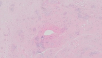
Sections reveal small amounts of residual atrophic thyroid follicles with many exhibiting oxyphilic metaplasia. There is focal squamous metaplasia present in some sections. The atrophic follicles are separated by mature dense hyaline fibrous tissue. Collagenous stroma contains scattered fibroblasts and a sprinkling of mature lymphocytes predominantly, with intermixed plasma cells and mononuclear cells. No lymphoid follicle or germinal center formation, eosinophilia, plasmacytosis, Reed-Sternberg cells, giant cell formation, calcifications or evidence of vascular invasion is present. The clinical presentation, gross findings and microscopic findings are interpreted as Hashimoto thyroiditis, fibrous atrophy variant.
Hashimoto thyroiditis is an autoimmune disease of the thyroid gland that usually presents clinically with hypothyroidism and has a high incidence in females. Histologic features of classical Hashimoto disease include prominent lymphocytic and plasmacytic infiltrates into the thyroid gland with lymphoid follicle formation, thyroid follicular atrophy, and oxyphilic metaplasia of thyroid folliclesIn approximately 10% of cases, usually ones that have markedly elevated autoantibody titers to thyroglobulin and thyroid peroxidase (>1:2500), the thyroid gland becomes more fibrotic. This occurs at a female:male ratio of approximately 9:1.
In the classical fibrous variant of Hashimoto thyroiditis, typical residual findings of prominent lymphoplasmacytic infiltrates with lymphoid germinal center formation are usually easily found in relation to prominent areas of fibrosis. In the fibrous atrophy variant, as seen here, usually the thyroid gland is smaller and exhibits extensive loss of thyroid follicles. Histologically, there is extensive fibrosis with sparse lymphoplasmacytic infiltrates, and marked thyroid follicular atrophy. In this case, there were no residual findings of prominent lymphoplasmacytic infiltrates, lymphoid follicle formation, or normal thyroid follicular architecture present in sections resulting in classification as fibrous atrophy variant.
The lymphocytic infiltrates are represented by both T- and B-lymphocytes. Quantitative studies of plasma cells reveal a predominance of IgG heavy chains and kappa light chains in Hashimoto thyroiditis. One study showed a 2:1 ratio of kappa light chains to lambda light chains of plasma cells in fibrous variant of Hashimoto thyroiditis. Another study revealed a subgroup of Hashimoto thyroiditis that contains IgG4 plasma cells. This subgroup exhibited progressively higher degree of fibrosis, more prominent lymphoplasmacytic infiltrates, a higher degree of thyroid follicular degeneration, and higher levels of autoantibodies.
Due to the clinical presentation of a firm to hard thyroid mass, it is not unusual to have these specimens or biopsies submitted for frozen section. The differential in these cases includes a malignant lymphoma, follicular carcinoma, and other carcinomas of the head and neck region invading into the thyroid gland. In many cases, squamous metaplasia is prominent in fibrous variant of Hashimoto thyroiditis resulting in additional challenges to rule out squamous carcinoma. Knowing the history that the patient is hypothyroid and has high titers of autoantibodies to thyroglobulin and thyroid peroxidase can be a strong clue that one is dealing with a variant of Hashimoto thyroiditis instead of a follicular carcinoma or squamous cell carcinoma invading the thyroid gland. Hashimoto thyroiditis has been associated with an increased incidence of lymphomas of the thyroid. Patients with Hashimoto thyroiditis have also been reported to have an increased incidence of papillary carcinoma and Hürthle cell carcinoma of the thyroid.
Amyloid deposition of the thyroid not associated with medullary carcinoma is a very rare event, and has been called amyloid goiter and is associated with systemic amyloidosis. When thyroid is involved with systemic amyloid, there is usually dysfunction of other organs that are involved. Amyloid of the thyroid exhibits a white to tan appearance and may appear waxy grossly. Histologically, there are amorphous eosinophilic extracellular deposits within interstitium, perivascular and vascular locations. There is usually foreign-body giant cell reaction present and calcifications may be identified, neither of which are present in this case. If amyloid is in the differential, a Congo red and/or thioflavin T stain would help differentiate the presence or absence of amyloid. If amyloid is found, it is wise to re-gross the resected thyroid tissue to rule out a hiding medullary carcinoma.
The possibility of a follicular carcinoma with Hürthle cell change or a Hürthle cell carcinoma must be considered in the differential. In view of the distorted follicles, some with Hürthle cell change, in a fibrous stroma might lead one to consider malignancy. The history of hypothyroidism with autoantibodies is supportive of Hashimoto thyroiditis. Features arguing against a Hürthle cell carcinoma include a lesion confined to the thyroid and which lacks vascular invasion or capsular invasion.
With the past history of Hodgkin lymphoma, the possibility of reoccurrence must be considered. Lymphocyte depleted classical Hodgkin lymphoma can exhibit a diffuse fibrosis pattern or a reticular pattern. In the diffuse fibrosis pattern, there is progressive fibrosis/sclerosis with rare Reed-Sternberg cells and a decrease in numbers of lymphocytes. In the reticular type, one sees prominent numbers of Reed-Sternberg cells, and Reed-Sternberg-like cells in a background of fibrosis with neutrophils, eosinophils, plasma cells, and histiocytes. Neither pattern is present in this case and no Reed-Sternberg cells or cells mimicking Reed-Sternberg cells are present. In a sclerosing lesion, nodular sclerosing Hodgkin lymphoma and other non-Hodgkin lymphomas might also be in the differential of a case of Hashimoto thyroiditis, fibrous variant. Multifocal sclerosing thyroiditis is a relatively new entity represented by multiple sites of fibrosclerosis throughout the thyroid gland with each exhibiting radial architecture.
In some cases, the number of foci are too numerous to count. At lower power, the lesion can mimic papillary microcarcinoma with small entrapped follicles. However, the nuclei lining the follicles typically lack optical clearing, nuclear grooves, and pseudoinclusions of papillary carcinoma of thyroid. Peri-fibrosclerotic thyroid tissue may exhibit follicular epithelial proliferation with hyperplastic changes, and in rare cases the lesions also contain foci with features of papillary microcarcinoma suggesting an evolution of hyperplasia to microcarcinoma. The pathogenesis is unknown but it has been compared to radial scars of the breast, some of which may also harbor small tubular carcinomas. Experts suggest that this thyroid lesion is initiated with an injury-producing scar followed by hyperplasia with possible evolution to carcinoma. This lesion has been found in the background of Hashimoto thyroiditis and in the normal thyroid.
In chronic (months to years afterward) post-radiation fibrosis, one would expect to see atypical fibroblasts within the fibrous/sclerosing reaction along with intimal arterial thickening due to fibroplasia and sclerosis. The presence of thyroid follicular nuclear atypia may also be seen along with other features that can mimic Hashimoto thyroiditis including thyroid follicular atrophy, oxyphilic change, squamous metaplasia, and lymphoplasmacytic infiltrates. In acute radiation changes, regional thyroid follicular hyperplasia, oxyphilic metaplasia, and lymphocytic infiltrates may be seen histologically.
Riedel thyroiditis is very rare and is seen in approximately 0.05 percent of thyroid resections, and it has also been known as Riedel struma, invasive fibrous thyroiditis, and ligneous thyroiditis after its gross "woody" appearance. Some feel that it is not a disease of the thyroid but a disorder that involves other head and neck structures and can be systemic including pancreas, biliary tract, mediastinum, and retroperitoneum. As a result, some favor classifying this entity as an inflammatory fibrosclerosing lesion or multifocal fibrosclerosis entity associated with IgG4 antibodies. It presents as a painless goiter and sometimes with dyspnea in adults with a female predominance, and patients are usually euthyroid. Physical exam reveals a stone-hard or woody thyroid gland on palpation. The process is bound to surrounding soft tissue. At surgery, the lesion is stone hard, difficult to section with a scalpel and contains dense fibrous bands extending outside of the thyroid into adjacent soft tissue and neck muscles resulting in complex surgical dissection. Histologically, there is extensive fibrosis of the thyroid gland containing keloid-like fibers associated with lymphocytes, plasma cells, including numerous IgA and IgG4 plasma cells, and eosinophils. Blood vessels and peripheral nerves are encased by fibrosclerosing stroma that extends into soft tissue, which is atypical for fibrosing variant of Hashimoto thyroiditis. There may be vasculitis with thrombosis present, which is also atypical for fibrosing variant of Hashimoto thyroiditis. Oxyphilic metaplasia of thyroid follicles typical of Hashimoto thyroiditis, fibrous variant, is absent in Riedel thyroiditis.
Supplementary Questions:
- Riedel thyroiditis is associated with IgG4-related inflammatory fibrosclerosing diseases.
- True
- False
- Patients with Hashimoto thyroiditis, fibrous variant, typically have higher autoantibodies to thyroid peroxidase and thyroglobulin than do patients with classical Hashimoto thyroiditis.
- True
- False
- Papillary microcarcinoma of thyroid can be found in association with multifocal sclerosing thyroiditis.
- True
- False
References
- Cheuk W, Chan JKC. IgG4-related sclerosing disease, a critical appraisal of an evolving clinicopathologic entity. Adv Anat Pathol. 2010;17(5):303-332.
- Heufelder AE, Goellner JR, Bahn RS, et al. Tissue eosinophilia and eosinophil degranulation in Riedel's invasive fibrous thyroiditis. J Clin Endocr Metab. 1996;81(3):977-984.
- Katz SM, Vickery AL. The fibrous variant of Hashimoto's thyroiditis. Hum Pathol. 1974;5(2):161-170.
- LiVolsi VA. The pathology of autoimmune thyroid disease: a review. Thyroid. 1994;4(3):333-339.
- Lloyd RV, Douglas BR, Young WF. Endocrine Diseases. Atlas of Nontumor Pathology,. first series, fascicle 1. Washington DC: Armed Forces Institute of Pathology; 2002:61-62.
- Mills SE, Carter D, Greenson JK, eds. Sternberg's Diagnostic Surgical Pathology. 5th ed. Philadelphia, PA: Lippincott Williams and Wilkins; 2010.
- Poli F, Trezzi R, Fellegara G, Rosai J. Multifocal sclerosing thyroiditis. Int J Surg Pathol. 2009;17:144.
- Rosai J. Rosai and Ackerman's Surgical Pathology. 10th ed. Philadelphia, PA: Mosby Elsevier; 2011.
Author
2014
Byron Crawford, MD
Surgical Pathology Committee
Tulane University School of Medicine
New Orleans, LA
Answer Key
- True (a).
- True (a).
- True (a).