- Home
- Member Resources
- Pathology Case Challenge
- Soft Tissue Left Thorax (Mediastinum)
Archive Case and Diagnosis
A 48-year-old woman presents with a 12.5 cm intrathoracic, mediastinal, well-circumscribed mass that is adhereent to the left uper lobe of the lung and near the 6th intercostal foramen. Radiologic heterogeneity is noted on chest CT with contrast.
Master List
- Cellular schwannoma
- Classic schwannoma
- Malignant peripheral nerve sheath tumor
- Neurofibroma
- Sarcomatoid carcinoma
- Solitary fibrous tumor, malignant
- Spindle cell melanoma
- Synovial sarcoma
Archive Case and Diagnosis
This case first appeared as Performance Improvement Program in Surgical Pathology (PIP) 2014, case 14 and is a cellular schwannoma.
Criteria for Diagnosis and Comments
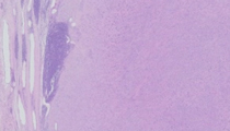
Cellular schwannoma is usually a centrally or axially located benign neoplasm that does not undergo malignant change. The tumor is often eccentric to nerve with a fusiform growth pattern and is entirely composed of Schwann cells. The histologic features of cystic degeneration, degenerative necrosis, cytologic atypia and increased mitoses are acceptable, yet marked pleomorphism, atypical mitoses or geographic necrosis associated with malignant neoplasms are not found. Reactive xanthoma cell inflammation is commonly observed, as part of central degeneration. The tumor is composed of cellular fascicular spindled areas, wavy and slightly plump nuclei, pink cytoplasm, and eosinophilic hyalinization of stroma and around vessels. The cellular area is designated “Antoni A” and it is without the well-formed Verocay bodies of classic schwannoma. The hypocellular myxoid (neurofibroma-like) areas of "Antoni B" are absent in cellular schwannoma. The current tumor also has a benign pseudoglandular appearance with attenuated gland-like epithelial-like cells that are thought to arise from pluripotential nerve sheath stem cells. Presence of a solitary mass in the retroperitoneum, thorax, abdomen or pelvis (central locations) may be observed. In the current case, the tumor arises from the Schwann cells of the intercostal nerve and extends into the thorax, pushing on the lower lobe of the lung.
Immunohistochemical stains on paraffin-embedded tissue demonstrate strong diffuse immunoreactivity for S100 protein (staining Schwann cells), focal GFAP and focal cross-reactivity with pankeratin. The tumor cells are negative for CD34, NFP, HMB45, SMA, and desmin. EMA stains the periphery (capsular area) of schwannoma, the perineurial or epineurial cells.
Classic schwannoma also arises from the Schwann cell eccentric to nerve, with a fusiform growth pattern, but with both cellular Antoni A regions with palisading Verocay bodies, and hypocellular Antoni B regions. These usually occur near peripheral, rather than central nerves. The immunophenotype is the same as for cellular schwannoma.
Synovial sarcoma can occur in an intrathoracic location. The monophasic form has scattered mast cells and also an eosinophilic stromal appearance, with staghorn hemangiopericytoid vasculature, ovoid cells, and denser cellularity. This can have a higher grade round cell appearance with perivascular tumor sparing and geographic necrosis, which is present focally (atypically) in the current lesion. Immunostains separate synovial sarcoma from schwannoma because the former are positive at least focally for EMA, pankeratin and CK7 and S100 protein usually only stains residual wavy neurons within tumor.
Malignant peripheral nerve sheath tumor is a high grade sarcoma with perivascular tumor sparing and geographic necrosis. It has tumor cells with high nuclear to cytoplasmic ratio imparting a low power “blue appearance” rather than the eosinophilic appearance typical of schwannoma. The tumor arises from nerve or neurofibroma, with invasion of surrounding structures or pseudocapsule, and is positive only focally for S100 protein, GFAP and usually negative for keratins, desmin, HMB45, and CD34.
Neurofibroma arises from nerve, so there is a centrally located entering and exiting nerve with a central more neural-like and peripheral more edematous appearance. The tumor cells are positive diffusely for S100 protein (Schwann cells), CD34 (intraneural CD34 positive fibroblasts) and there are residual NFP neurites in the center. GFAP is more often negative in tumor cells than in schwannoma. EMA stains the periphery of neurofibroma when it is still intraneural.
Sarcomatoid carcinoma can occur in this region and be either primary pulmonary adenocarcinoma (CK18 and TTF1 positive) or squamous cell carcinoma (CK5/6 positive). Sarcomatoid carcinoma demonstrates marked malignant morphologic features as well as at least focal presence of keratins.
Solitary fibrous tumor may be present in this thoracic location and can have a similar eosinophilic appearance with haphazard plump cells, and scant mitotic activity. When centrally located and greater than 5.0 cm in diameter, with mitoses greater than 4/10 hpf, increased cytologic atypia, hemorrhage and necrosis, these can be considered low grade malignant neoplasms. CD34 would be diffusely positive, and S100 protein would be negative.
Spindle cell melanoma can occur in this location in thorax, often as a metastasis from a skin primary. This can be difficult to separate from schwannoma, especially since these lesions are both spindled, eosinophilic and diffusely strongly positive for S100 protein. Melanoma would have prominent cherry red nucleoli and/or intranuclear cytoplasmic inclusions and often atypical mitotic activity, marked cytologic atypia and geographic necrosis. Melanoma markers, including HMB45, Melan-A, tyrosinase and MiTf (microphthalmia transcription factor) are often negative in spindle cell melanoma. The more bland appearance, absence of mitotic activity and xanthomatous degeneration of the current lesion favors schwannoma over spindle cell melanoma.
Supplementary Questions:
- Cellular schwannoma can be separated from classic schwannoma by its central or axial (proximal) location and its predominance of Antoni A region, absence of Verocay body, and lack of Antoni B region.
- True
- False
- Which of the following is the hardest tumor to separate from schwannoma because of diffuse, strong S100 protein reactivity in both lesions?
- Malignant peripheral nerve sheath tumor
- Neurofibroma
- Sarcomatoid carcinoma
- Spindle cell melanoma
- Neurofibroma and schwannoma share their S100, CD34, EMA, GFAP, and keratin staining patterns.
- True
- False
References
- Bernaba BN, Vogiatzis PI, Binder SW, Cassarino DS. Potentially useful markers for desmoplastic melanoma: an analysis of KBA.62, p-AKT, and ezrin. Am J Dermatopathol. 2011;33(4):333-337.
- Fanburg-Smith JC, Majidi M, Miettinen M. Keratin expression in schwannoma; a study of 115 retroperitoneal and 22 peripheral schwannomas. Mod Pathol. 2006;19(1):115-121.
- Kao GF, Laskin WB, Olsen TG. Solitary cutaneous plexiform neurilemmoma (schwannoma): a clinicopathologic, immunohistochemical, and ultrastructural study of 11 cases. Mod Pathol. 1989;2(1):20-26.
- Michal M, Fanburg-Smith JC, Mentzel T, et al. Dendritic cell neurofibroma with pseudorosettes: a report of 18 cases of a distinct and hitherto unrecognized neurofibroma variant. Am J Surg Pathol. 2001;25(5):587-594.
- Paredes I, Jimenez Roldán L, Ramos A, Lobato RD, Ricoy JR. Intraparenchymal schwannomas: report of two new cases studied with MRI and review of the literature. Clin Neurol Neurosurg. 2012;114(1):42-46.
- Schulman FY, Johnson TO, Facemire PR, Fanburg-Smith JC. Feline peripheral nerve sheath tumors: histologic, immunohistochemical, and clinicopathologic correlation (59 tumors in 53 cats). Vet Pathol. 2009;46(6):1166-1180.
Author
2014
Julie C. Fanburg-Smith, MD FCAP
Surgical Pathology Committee
Sibley Memorial Hospital of Johns Hopkins Medicine
Department of Pathology and Surgical Pathology Consultation Services
Washington DC
Answer Key
- True (a).
- Spindle cell melanoma (d).
- False (b).