- Home
- Member Resources
- Pathology Case Challenge
- Left Atrium, Heart
Clinical Summary
A 53-year-old man with clinical history of hypertension and hypercholesterolemia is admitted to the emergency room due to a possible stroke. A transthoracic echocardiogram is unremarkable. On further evaluation, a transesophageal echocardiogram reveals an oval and pedunculated mass, measuring 2.0 cm, adherent to the left atrial wall. The mass is mobile and distensible, protruding to the left atrial appendage during atrial systole. Due to its embolic potential, the mass is removed. The resected mass is soft, polypoid, and pedunculated, with a gelatinous cut surface and focal dark red areas on the periphery.
Master List
- Cardiac (atrial) myxoma
- Metastatic mucinous carcinoma/sarcoma
- Neurofibroma
- Nodular fasciitis
- Organizing thrombus
- Papillary fibroelastoma
Archive Case and Diagnosis
This case first appeared as Performance Improvement Program in Surgical Pathology (PIP) 2014, case 06 and is a cardiac (atrial) myxoma.
Criteria for Diagnosis and Comments
Cardiac myxoma is the most common heart tumor. Over 90% of cases originate in the atria, the majority in left atrium. It typically originates from the atrial septum near the fossa ovale, but other sites of attachment have been reported including ventricle and cardiac valves. Multiple myxomas are uncommon and may be part of the myxoma syndrome.
Cardiac myxomas are either pedunculated or sessile but mobile. The tumor usually presents as a polypoid mass with multiple fronds or as globular masses with a smooth surface. They are usually soft and friable with a distinctive gelatinous appearance and spotty hemorrhage on the surface. Some myxomas have an overall firm consistency. Occasionally gross calcifications can be observed, a condition also known as "petrified cardiac myxoma." The cut surface of a cardiac myxoma shows a gelatinous and shiny appearance. The peripheral aspects usually appear dark and hemorrhagic. Of note, multiple sections should be taken from different areas to ensure the evaluation of its overall histopathologic features.
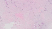
Histopathologically, the overall picture of cardiac myxomas is composed of a myxomatous matrix and a dispersed cellular component.
The neoplastic cells (myxoma cell) may appear elongated and fusiform, polyhedral, or stellate. The cytoplasm is mostly homogeneous, sometimes finely vacuolated, and is usually slightly eosinophilic. The nucleus may be elongated, rounded, or oval and may vary from pale to intensely hyperchromatic. Mitoses are virtually absent. The myxoma cells are arranged in various patterns. The common pattern is a ring-like arrangement with single or multiple layers of cells surrounding vascular channels. They may also appear as complex interlacing networks or single strands. The clusters of myxoma cells are often associated with an extensive halo of myxoid extracellular matrix, in contrast to the surrounding matrix that is more condensed and stains slightly eosinophilic. Other possible arrangements of myxoma cells include the formation of small nests and single cells dispersed throughout the myxoid stroma.
Myxoma cells with an appearance of multinucleated cells can also be present. The surface of the tumor may be covered by a single layer of endothelial cells. In approximately 5% of all cardiac myxomas gland-like structures lined by flat to cuboidal or columnar cells are seen. These cells are positive with alcian blue, mucicarmine, and periodic acid–Schiff (PAS) (diastase-resistant) and show positive immunoreactivity for cytokeratins, raising the differential diagnosis of metastatic carcinoma with mucinous features. Myxoma cells usually stain positively for vimentin, but do not stain with endothelial markers. Overall, the appearance of the myxoma cells is highly variable even within the same tumor. Therefore, the overall picture is much more important to render a correct diagnosis than the cytology of a few cells. Other cell types frequently observed in cardiac myxomas are macrophages, lymphocytes, and plasma cells. These cells are more frequently observed in the base of the tumor and can also be found in the adjacent myocardium. Mast cells and foci of extramedullary hematopoiesis are occasionally seen.
The myxoid stroma stains strongly with alcian blue, unaffected by digestion with hyaluronidase, and may stain patchily with mucicarmine and PAS (diastase-resistant). It contains, variably, connective tissue fibers with features of reticulin, collagen, and elastin. Fibrous tissue is most noted in the stalk of the tumor, a site often dominated by the presence of large, thick-walled arteries. Hemorrhages within the stroma are a common finding in myxomas. In addition, microscopic foci of calcification can be noted. Occasionally the dystrophic calcifications within the tumor are so extensive that the tumor is almost completely transformed to a calcified mass.
The cardiac myxoma, especially the multiple and familial form, may present as manifestations of Carney complex. This peculiar syndrome is known by a variety of names, including "Swiss syndrome" and Carney syndrome, as well as the acronyms NAME syndrome (nevi, atrial myxoma, myxoid neurofibroma and neurofibromata, and ephelides) and LAMB syndrome (lentigines, atrial myxoma, mucocutaneous myxoma, blue nevi). The syndrome is also known as "familial endocrine myxolentiginosis," which is descriptive of the major components of the syndrome. This syndrome consists of a variable complex of mucocutaneous, visceral, and endocrine disorders, not all of which are necessarily present in a single patient. Those lesions include cardiac myxomas (with a strong tendency to multiplicity), cutaneous myxomas (single or multiple), mammary myxoid fibroadenomas (single or multiple), spotty mucocutaneous pigmentation (including lentigines and blue nevi and combinations), psammomatous melanotic schwannomas, primary pigmented nodular adrenocortical disease (including patients who are symptomatic with Cushing syndrome), testicular tumors (characteristically Sertoli cell tumors, usually bilateral and multicentric), and pituitary growth hormone-secreting tumors (which may cause acromegaly or gigantism). If myxomas, spotty pigmentation, and endocrine overactivity are present in three successive generations of one family, the first-degree relatives of affected persons should be thoroughly examined for this syndrome. The syndrome is inherited in an autosomal dominant fashion and most cases result from mutations in the PRKAR1A gene resulting in increased activity of protein kinase A. Some individuals with the complex have an undefined abnormality at chromosome 2p16.
As mentioned above, cardiac myxomas with gland-like spaces may elicit the suspicion of a metastatic mucinous carcinoma. The gland-like cells may appear as mucus-secreting cells and immunohistochemical analysis reveals positivity for cytokeratins. However, these cells are cytologically bland, and usually focal. Therefore, it is important to evaluate the entire tumor and obtain detailed clinical information to rule out this possibility.
Other important tumors to be considered in the differential diagnosis are the myxoid variants of soft tissue sarcomas such as myxoid malignant fibrous histiocytoma (myxofibrosarcoma) and liposarcoma. Most cardiac sarcomas are also found in the left atrium and often have myxoid stroma.
Careful examination can identify foci of atypical spindle cells with nuclear pleomorphism, hypercellularity, and particularly important, increased mitotic activity. The sarcoma cells often exhibit storiform growth pattern with arborizing vessels, and do not form cords or ring structures around blood vessels.
Neurofibromas and nodular fasciitis in the heart are extremely rare, but the histopathologic features are the same as those encountered in the soft tissue. Neurofibromas may present as parts of neurofibromatosis type I (von Recklinghausen disease). The condition may lead to right ventricular outflow tract obstruction.
Organized thrombus may appear as a hypocellular fibrous endocardial mass with myxoid background. However, the spindle mesenchymal cells are not arranged in cords, nests, or ring structures. The base of endocardial lesions does not contain the lymphocytic infiltrates or prominent thick-walled vessels that are often present in myxoma.
Although papillary fibroelastoma is a relatively rare lesion, it ranks third among the primary benign heart tumors in adults. These papillary tumors can involve any of the heart valves and may be an incidental finding. The left-sided lesions are more likely to be symptomatic, and location on the aortic valve is potentially hazardous. These tumors consist of a cluster of filiform papillae attached to the endocardium, either sessile or connected by a distinct but short pedicle. The tumors most frequently arise on the valves, mainly the aortic valve. Occasionally, a patient may develop multiple fibroelastomas. Histologically, the tumor consists of a central core of dense, almost acellular collagen, occasionally surrounded by a myxomatous matrix. The peripheral rim often contains coarse and fragmented elastin fibrils. The surface lining is a layer of hyperplastic endothelial cells.
Supplementary Questions:
- Which of the following is the most common primary cardiac tumor?
- Cardiac fibroma
- Cardiac myxoma
- Cardiac rhabdomyoma
- Cardiac teratoma
- Papillary fibroelastoma
- Which systemic abnormality maybe associated with cardiac myxoma?
- Carney complex (Carney syndrome)
- Carney triad
- Marfan syndrome
- Neurofibromatosis type I
- Neurofibromatosis type II
- Which abnormality is not present in Carney complex (Carney syndrome)?
- Cardiac myxoma
- Cushing syndrome
- Myxoid neurofibroma
- Pulmonary chondroma
- Spotty skin pigmentation
References
- Acebo E, Val-Bernal JF, Gomez-Roman JJ, Revuelta JM. Clinicopathologic study and DNA analysis of 37 cardiac myxomas: A 28-year experience. Chest. 2003;123:1379-1385.
- Fletcher CD. Diagnostic Histopathology of Tumors. 3rd ed. Philadelphia, PA: Elsevier Limited; 2007;7-40.
- Gattuso P, Reddy VB, David O, Spitz DJ, Haber MH. Differential Diagnosis in Surgical Pathology. 2nd ed. Philadelphia, PA: Saunders; 2010;949-968.
- Swartz MF, Lutz CJ, Chandan VS, Landas S, Fink GW. Atrial myxomas: pathologic types, tumor location, and presenting symptoms. J Card Surg. 2006;21(4):435-440.
Authors
2014
Zhiyong Ren, MD
Pathology Resident
University of Alabama at Birmingham
Birmingham, AL
Thomas S. Winokur, MD
Surgical Pathology Committee
Department of Pathology
University of Alabama at Birmingham
Birmingham, AL
Answer Key
- Cardiac myxoma (b).
- Carney complex (Carney syndrome) (a).
- Pulmonary chondroma (d).