- Home
- Digital Pathology Resource Guide
- Background and Framework
Background and Framework
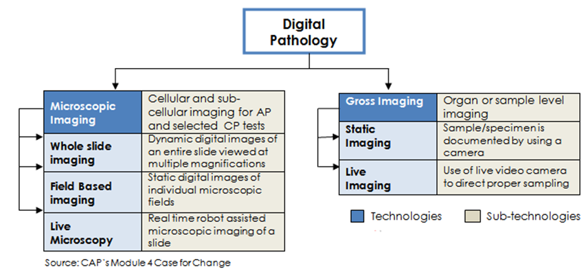
This Resource Guide is focused on microscopic imaging (primarily Whole Slide Imaging (WSI)) within digital pathology; however, gross imaging is also part of digital pathology.
Suggested Articles and Resources
- A) Whole-slide Imaging: Routine Pathologic Diagnosis
Cornish TC, Swapp RE, Kaplan KJ. Whole-slide Imaging: Routine Pathologic Diagnosis. Adv Anat Pathol 2012;19(3):152-159. doi: 10.1097/PAP.0b013e318253459e. Full text available from Advances in Anatomic Pathology (subscription required)
PMID: 22498580
NOTE: Also cited in Section 7.8 - B) Whole Slide Imaging in Pathology: Advantages, Limitations, and Emerging Perspectives
Farahani N, Parwani AV, Pantanowitz L. Whole slide imaging in pathology: advantages, limitations, and emerging perspectives. Pathology and Laboratory Medicine International. 2015(7): 23-33.
Summary: Significant technologic gains have led to the adoption of innovative digital imaging solutions in pathology. Whole slide imaging (WSI), which refers to scanning of conventional glass slides in order to produce digital slides, is the most recent imaging modality being employed by pathology departments worldwide. WSI continues to gain traction among pathologists for diagnostic, educational, and research purposes. This article provides a technologic review of WSI platforms and covers clinical and nonclinical pathology applications of these imaging systems. Barriers to adoption of WSI include limiting technology, image quality, problems with scanning all materials (eg, cytology slides), cost, digital slide storage, inability to handle high-throughput routine work, regulatory barriers, ergonomics, and pathologists' reluctance. Emerging issues related to clinical validation, standardization, and forthcoming advances in the field are also addressed.
Free full text available from Pathology and Laboratory Medicine International
Background and Framework
This Resource Guide is focused on microscopic imaging (primarily Whole Slide Imaging (WSI)) within digital pathology; however, gross imaging is also part of digital pathology.
Suggested Articles and Resources
- A) Whole-slide Imaging: Routine Pathologic Diagnosis
Cornish TC, Swapp RE, Kaplan KJ. Whole-slide Imaging: Routine Pathologic Diagnosis. Adv Anat Pathol 2012;19(3):152-159. doi: 10.1097/PAP.0b013e318253459e. Full text available from Advances in Anatomic Pathology (subscription required)
PMID: 22498580
NOTE: Also cited in Section 7.8 - B) Whole Slide Imaging in Pathology: Advantages, Limitations, and Emerging Perspectives
Farahani N, Parwani AV, Pantanowitz L. Whole slide imaging in pathology: advantages, limitations, and emerging perspectives. Pathology and Laboratory Medicine International. 2015(7): 23-33.
Summary: Significant technologic gains have led to the adoption of innovative digital imaging solutions in pathology. Whole slide imaging (WSI), which refers to scanning of conventional glass slides in order to produce digital slides, is the most recent imaging modality being employed by pathology departments worldwide. WSI continues to gain traction among pathologists for diagnostic, educational, and research purposes. This article provides a technologic review of WSI platforms and covers clinical and nonclinical pathology applications of these imaging systems. Barriers to adoption of WSI include limiting technology, image quality, problems with scanning all materials (eg, cytology slides), cost, digital slide storage, inability to handle high-throughput routine work, regulatory barriers, ergonomics, and pathologists' reluctance. Emerging issues related to clinical validation, standardization, and forthcoming advances in the field are also addressed.
Free full text available from Pathology and Laboratory Medicine International